Hase T
J Bacteriol. 1983 May;154(2):879-92. doi: 10.1128/jb.154.2.879-892.1983.
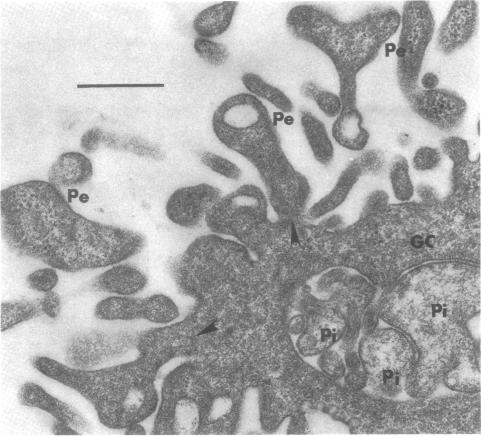
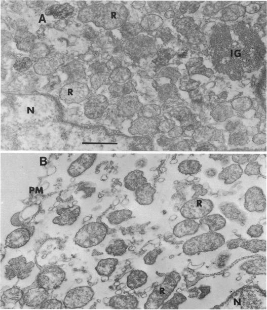
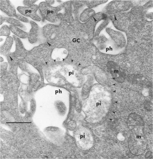
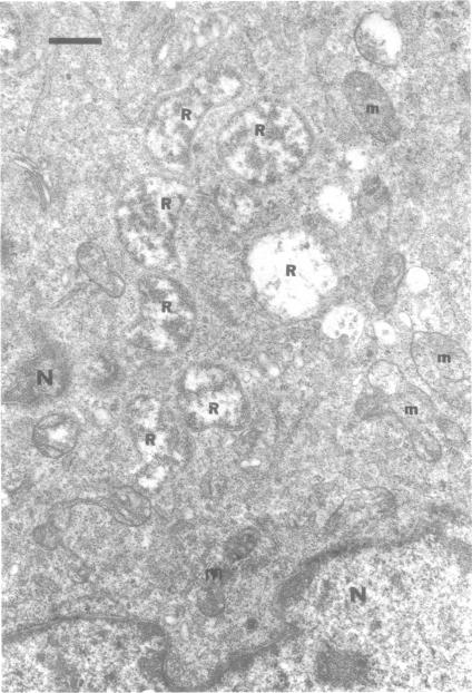
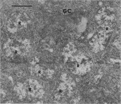
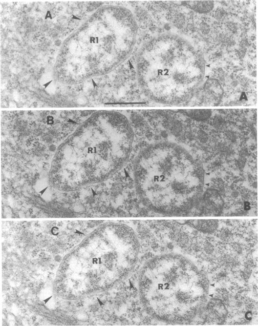
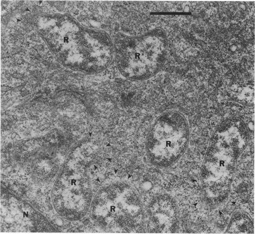
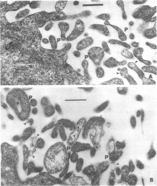
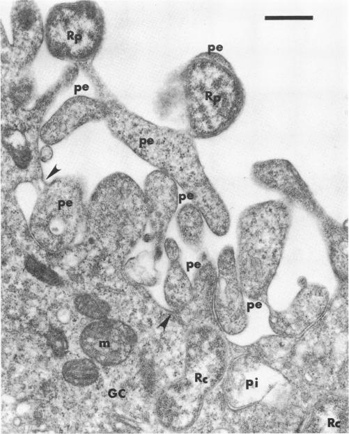
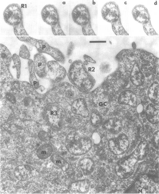
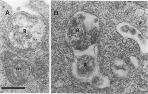
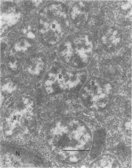
Irradiated L cells infected with Rickettsia tsutsugamushi were studied under the electron microscope to define the morphological growth pattern of the organism. For 2 days after inoculation, no rickettsiae were found either extra- or intracellularly; after 2 days multiple rickettsiae appeared within the host cells without morphological evidence of their entry. These observations showed that the rickettsiae within the cell were assembled in situ by segregation of portions of the granular cytoplasm and subsequent internal differentiation and surface membrane assembly of the segregated bodies. The protoplasmic (P) bodies, which seemed to be formed by shedding infected-cell granular cytoplasm, consistently appeared on the surface and within the phagosomes of the host cells. Rickettsiae were occasionally seen entering host cells in the later phase of infection; these were apparently the ones assembled within the P bodies. This suggested that the P bodies, and not the rickettsiae, were the major infectious particles that transmitted the rickettsial genetic substance among the host cells. On the basis of the present morphological observations, viral-type multiplication for R. tsutsugamushi is proposed.
用电子显微镜研究了感染恙虫病立克次体的受辐照L细胞,以确定该生物体的形态生长模式。接种后2天,细胞内外均未发现立克次体;2天后,宿主细胞内出现多个立克次体,且无其进入的形态学证据。这些观察结果表明,细胞内的立克次体是通过颗粒状细胞质部分的分离以及随后分离体的内部分化和表面膜组装而在原位组装的。原生质(P)体似乎是通过脱落受感染细胞的颗粒状细胞质形成的,始终出现在宿主细胞的表面和吞噬体内。在感染后期偶尔可见立克次体进入宿主细胞;这些显然是在P体内组装的立克次体。这表明P体而非立克次体是在宿主细胞间传递立克次体遗传物质的主要感染性颗粒。基于目前的形态学观察结果,提出了恙虫病立克次体的病毒型增殖方式。