• 文献检索
  • 文档翻译
  • 深度研究
  • 学术资讯
  • Suppr Zotero 插件Zotero 插件
  • 邀请有礼
  • 套餐&价格
  • 历史记录
应用&插件
Suppr Zotero 插件Zotero 插件浏览器插件Mac 客户端Windows 客户端微信小程序
定价
高级版会员购买积分包购买API积分包
服务
文献检索文档翻译深度研究API 文档MCP 服务
关于我们
关于 Suppr公司介绍联系我们用户协议隐私条款
关注我们

Suppr 超能文献

核心技术专利:CN118964589B侵权必究
粤ICP备2023148730 号-1Suppr @ 2026

文献检索

告别复杂PubMed语法,用中文像聊天一样搜索,搜遍4000万医学文献。AI智能推荐,让科研检索更轻松。

立即免费搜索

文件翻译

保留排版,准确专业,支持PDF/Word/PPT等文件格式,支持 12+语言互译。

免费翻译文档

深度研究

AI帮你快速写综述,25分钟生成高质量综述,智能提取关键信息,辅助科研写作。

立即免费体验

聚球藻属菌株CLII规则构建的表层的三维结构。

Three-dimensional structure of the regularly constructed surface layer from Synechocystis sp. strain CLII.

作者信息

Karlsson B, Vaara T, Lounatmaa K, Gyllenberg H

出版信息

J Bacteriol. 1983 Dec;156(3):1338-43. doi: 10.1128/jb.156.3.1338-1343.1983.

DOI:10.1128/jb.156.3.1338-1343.1983
PMID:6417112
原文链接:https://pmc.ncbi.nlm.nih.gov/articles/PMC217985/
Abstract

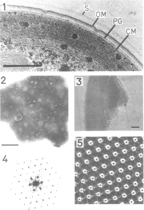
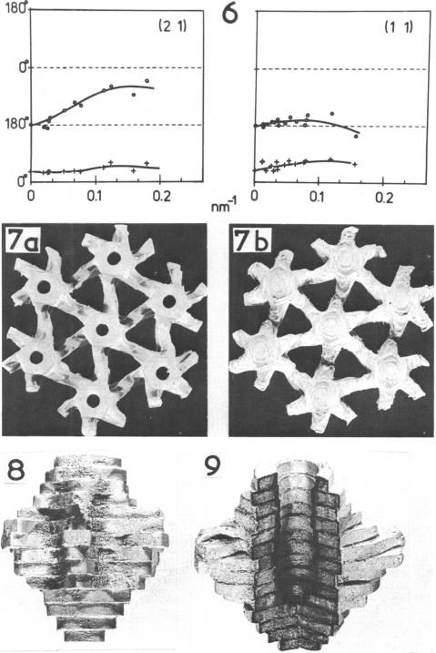
The isolated, outermost cell wall layer from Synechocystis sp. strain CLII is described using electron microscopy and Fourier reconstruction to study the three-dimensional structure of the proteins within the layer to a resolution of ca. 3 nm. This surface layer forms regular hexagonal arrays (a = b = 15.2 nm). The two-dimensional space group is p6. The monomer proteins form hexamers arranged around a central hollow cylinder. The linkers between the hexamers are of the delta type and are located approximately in the central section between the top and bottom of the protein layer.

摘要

使用电子显微镜和傅里叶重建技术,对来自聚球藻属菌株CLII的分离出的最外层细胞壁层进行了描述,以研究该层内蛋白质的三维结构,分辨率约为3纳米。该表面层形成规则的六边形阵列(a = b = 15.2纳米)。二维空间群为p6。单体蛋白形成围绕中心空心圆柱体排列的六聚体。六聚体之间的连接体为δ型,大致位于蛋白质层顶部和底部之间的中心部分。

https://cdn.ncbi.nlm.nih.gov/pmc/blobs/28cc/217985/146ebbd2f035/jbacter00241-0371-a.jpg
https://cdn.ncbi.nlm.nih.gov/pmc/blobs/28cc/217985/4ee7b4ac41eb/jbacter00241-0370-a.jpg
https://cdn.ncbi.nlm.nih.gov/pmc/blobs/28cc/217985/146ebbd2f035/jbacter00241-0371-a.jpg
https://cdn.ncbi.nlm.nih.gov/pmc/blobs/28cc/217985/4ee7b4ac41eb/jbacter00241-0370-a.jpg
https://cdn.ncbi.nlm.nih.gov/pmc/blobs/28cc/217985/146ebbd2f035/jbacter00241-0371-a.jpg

相似文献

1
Three-dimensional structure of the regularly constructed surface layer from Synechocystis sp. strain CLII.聚球藻属菌株CLII规则构建的表层的三维结构。
J Bacteriol. 1983 Dec;156(3):1338-43. doi: 10.1128/jb.156.3.1338-1343.1983.
2
Ultrastructure of the cell wall of a Synechocystis strain.聚球藻属菌株细胞壁的超微结构
Can J Microbiol. 1980 Feb;26(2):204-8. doi: 10.1139/m80-031.
3
S-layers on cell walls of cyanobacteria.蓝细菌细胞壁上的表面层
Micron. 2002;33(3):257-77. doi: 10.1016/s0968-4328(01)00031-2.
4
[Fine structure of the cell wall in various Cyanophyceae].
Zentralbl Bakteriol Orig A. 1974;228(1):189-92.
5
Carotenoid-containing outer membrane of Synechocystis sp. strain PCC6714.集胞藻PCC6714株含类胡萝卜素的外膜
J Bacteriol. 1985 Oct;164(1):384-9. doi: 10.1128/jb.164.1.384-389.1985.
6
Fine structure of the cell wall of the cyanobacterium Microcoleus vaginatus (Vauch.) Gomont.鞘丝藻(Vauch.)Gomont的细胞壁精细结构
Cytobios. 1981;30(117):19-27.
7
Three-dimensional structure of the regular surface layer (HPI layer) of Deinococcus radiodurans.耐辐射球菌规则表面层(HPI层)的三维结构。
J Mol Biol. 1986 Jan 20;187(2):241-50. doi: 10.1016/0022-2836(86)90231-7.
8
Surface arrays on the cell wall of Spirillum metamorphum.变形螺菌细胞壁上的表面阵列。
J Bacteriol. 1975 Dec;124(3):1529-44. doi: 10.1128/jb.124.3.1529-1544.1975.
9
Isolation of the carotenoid-containing cell wall of three unicellular cyanobacteria.三种单细胞蓝藻含类胡萝卜素细胞壁的分离
J Bacteriol. 1983 Jul;155(1):345-50. doi: 10.1128/jb.155.1.345-350.1983.
10
Phycobilisomes in spheroplasts of Anacystis nidulans.
J Bacteriol. 1978 Sep;135(3):1137-40. doi: 10.1128/jb.135.3.1137-1140.1978.

引用本文的文献

1
The sll1951 gene encodes the surface layer protein of Synechocystis sp. strain PCC 6803.sll1951 基因编码集胞藻 PCC 6803 菌株的表面层蛋白。
J Bacteriol. 2013 Dec;195(23):5370-80. doi: 10.1128/JB.00615-13. Epub 2013 Sep 27.
2
Fatty acid production in genetically modified cyanobacteria.在转基因蓝藻中产生脂肪酸。
Proc Natl Acad Sci U S A. 2011 Apr 26;108(17):6899-904. doi: 10.1073/pnas.1103014108. Epub 2011 Apr 11.
3
Cyanobacterial cell walls: news from an unusual prokaryotic envelope.蓝细菌细胞壁:来自一种独特原核生物包膜的新情况

本文引用的文献

1
Two improved methods for obtaining axenic cultures of cyanobacteria.两种改进的获得蓝藻无菌培养物的方法。
Appl Environ Microbiol. 1979 Nov;38(5):1011-4. doi: 10.1128/aem.38.5.1011-1014.1979.
2
Structure of the regular surface layer of Sporosarcina ureae.尿素芽孢杆菌规则表面层的结构
J Bacteriol. 1980 Apr;142(1):302-9. doi: 10.1128/jb.142.1.302-309.1980.
3
Structure of the regular surface layer of Spirillum putridiconchylium.腐败螺菌规则表面层的结构
J Bacteriol. 2000 Mar;182(5):1191-9. doi: 10.1128/JB.182.5.1191-1199.2000.
4
Structures of two different surface layers found in six Bacteroides strains.在六种拟杆菌菌株中发现的两种不同表层结构。
J Bacteriol. 1985 Dec;164(3):1278-82. doi: 10.1128/jb.164.3.1278-1282.1985.
5
Structure of the Azotobacter vinelandii surface layer.棕色固氮菌表层的结构。
J Bacteriol. 1987 Feb;169(2):802-10. doi: 10.1128/jb.169.2.802-810.1987.
6
Three-dimensional structure of the tetragonal surface layer of Sporosarcina ureae.尿素芽孢杆菌四方表面层的三维结构。
J Bacteriol. 1986 Oct;168(1):309-17. doi: 10.1128/jb.168.1.309-317.1986.
7
Three-dimensional structure of the T-layer of Bacillus sphaericus P-1.
J Bacteriol. 1986 Oct;168(1):303-8. doi: 10.1128/jb.168.1.303-308.1986.
8
Three-dimensional structure of the surface layer protein of Aquaspirillum serpens VHA determined by electron crystallography.
J Bacteriol. 1986 Sep;167(3):1025-34. doi: 10.1128/jb.167.3.1025-1034.1986.
9
Microbiological aspects of Helicobacter pylori (Campylobacter pylori).幽门螺杆菌(幽门弯曲菌)的微生物学特性
Eur J Clin Microbiol Infect Dis. 1990 Jan;9(1):1-13. doi: 10.1007/BF01969526.
10
Participation of a cyanobacterial S layer in fine-grain mineral formation.蓝细菌S层在细粒矿物形成中的作用。
J Bacteriol. 1992 Dec;174(24):7971-81. doi: 10.1128/jb.174.24.7971-7981.1992.
J Mol Biol. 1980 Feb 15;137(1):1-8. doi: 10.1016/0022-2836(80)90153-9.
4
Purification and disposition of a surface protein associated with virulence of Aeromonas salmonicida.与杀鲑气单胞菌毒力相关的一种表面蛋白的纯化与特性分析
J Bacteriol. 1981 Sep;147(3):1077-84. doi: 10.1128/jb.147.3.1077-1084.1981.
5
Structural studies of the outer envelope of Chlamydia trachomatis by electron microscopy.通过电子显微镜对沙眼衣原体外部包膜进行的结构研究。
J Mol Biol. 1982 Nov 15;161(4):579-90. doi: 10.1016/0022-2836(82)90409-0.
6
The correlation averaging of a regularly arranged bacterial cell envelope protein.规则排列的细菌细胞包膜蛋白的相关性平均
J Microsc. 1982 Aug;127(Pt 2):127-38. doi: 10.1111/j.1365-2818.1982.tb00405.x.
7
Structure of the regular surface layer of Aquaspirillum serpens MW5.水螺菌MW5规则表面层的结构
J Bacteriol. 1982 Apr;150(1):348-57. doi: 10.1128/jb.150.1.348-357.1982.
8
Cell wall proteins of Aquaspirillum serpens.蜿蜒水螺菌的细胞壁蛋白
J Bacteriol. 1981 Jun;146(3):1083-90. doi: 10.1128/jb.146.3.1083-1090.1981.
9
Ultrastructure of the cell wall of a Synechocystis strain.聚球藻属菌株细胞壁的超微结构
Can J Microbiol. 1980 Feb;26(2):204-8. doi: 10.1139/m80-031.
10
Three-dimensional structure of ubiquinol:cytochrome c reductase from Neurospora mitochondria determined by electron microscopy of membrane crystals.通过膜晶体电子显微镜确定的来自粗糙脉孢菌线粒体的泛醇:细胞色素c还原酶的三维结构。
J Mol Biol. 1981 Jun 25;149(2):259-74. doi: 10.1016/0022-2836(81)90301-6.