Tachibana M
J Physiol. 1983 Dec;345:329-51. doi: 10.1113/jphysiol.1983.sp014981.
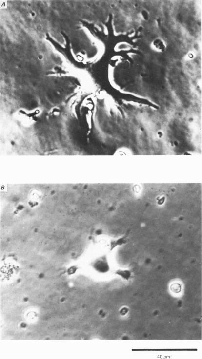
Solitary horizontal cells, dissociated from papain-treated goldfish retinas, produce action potentials and show a non-linear current-voltage relationship. Underlying ion-conductance mechanisms were analysed by a single-micro-electrode voltage-clamp technique. Pharmacological and ion-substitution experiments revealed that ionic currents could be separated into at least four voltage-dependent currents: a Ca current and three types of K currents. The Ca current was activated by membrane depolarization beyond -45 mV, reached a maximal value near 0 mV, and became smaller at more positive potentials. By extrapolation, the reversal potential was estimated to be approximately +50 mV. The Ca current was inactivated by accumulation of intracellular Ca ions but not by membrane depolarization. Co ions (4mM) blocked this current. The first type of K current showed anomalous (inward-going) rectification near the resting potential (congruent to -60 mV). Hyperpolarization from the resting level produced a large, almost steady inward current, while depolarization evoked only a small, steady outward current. The current-voltage relationship revealed a shallow negative resistance region at membrane potentials beyond -50 mV. The current was blocked by Cs (10 mM) or Ba (1 mM) ions. The second type of K current (the transient outward current) was activated by membrane depolarization beyond -25 mV. The peak amplitude increased almost exponentially as the membrane was depolarized. During steady depolarization this current decayed exponentially (time constant congruent to 500 ms at +20 mV). The current was inactivated by conditioning depolarization (greater than 10 s) beyond -30 mV and blocked by 4-aminopyridine (10 mM). The third type of K current was the maintained outward current which was activated by membrane depolarization beyond -20 mV, increased to a steady level in a few hundred milliseconds, and showed little inactivation. The amplitude increased as the membrane was depolarized. The current was blocked by tetraethylammonium ions (20 mM). A Ca-mediated K current was not detected. Action potentials and the non-linear current-voltage relationship of solitary horizontal cells can be explained qualitatively by the combination of the four ionic currents.
从木瓜蛋白酶处理过的金鱼视网膜中分离出的单个水平细胞能产生动作电位,并呈现出非线性电流-电压关系。通过单微电极电压钳技术分析其潜在的离子电导机制。药理学和离子替代实验表明,离子电流可至少分为四种电压依赖性电流:一种钙电流和三种钾电流。钙电流在膜去极化超过 -45 mV 时被激活,在接近 0 mV 时达到最大值,在更正的电位时变小。通过外推法,反转电位估计约为 +50 mV。钙电流因细胞内钙离子的积累而失活,但不因膜去极化而失活。钴离子(4 mM)可阻断该电流。第一种钾电流在静息电位(约 -60 mV)附近表现出反常(内向)整流。从静息水平超极化会产生一个大的、几乎稳定的内向电流,而去极化仅诱发一个小的、稳定的外向电流。电流-电压关系在膜电位超过 -50 mV 时显示出一个浅的负电阻区域。该电流被铯(10 mM)或钡(1 mM)离子阻断。第二种钾电流(瞬时外向电流)在膜去极化超过 -25 mV 时被激活。峰值幅度随着膜去极化几乎呈指数增加。在持续去极化期间,该电流呈指数衰减(在 +20 mV 时时间常数约为 500 ms)。该电流因超过 -30 mV 的预处理去极化(大于 10 s)而失活,并被 4-氨基吡啶(10 mM)阻断。第三种钾电流是持续外向电流,在膜去极化超过 -20 mV 时被激活,在几百毫秒内增加到稳定水平,且几乎没有失活。幅度随着膜去极化而增加。该电流被四乙铵离子(20 mM)阻断。未检测到钙介导的钾电流。单个水平细胞的动作电位和非线性电流-电压关系可以通过这四种离子电流的组合进行定性解释。