Franson T R, Sheth N K, Rose H D, Sohnle P G
J Clin Microbiol. 1984 Sep;20(3):500-5. doi: 10.1128/jcm.20.3.500-505.1984.
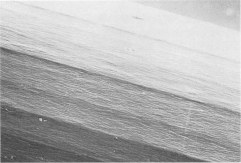
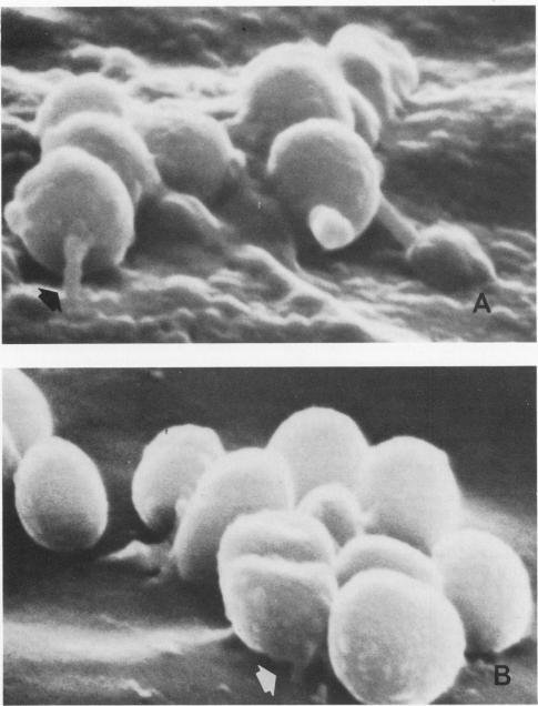
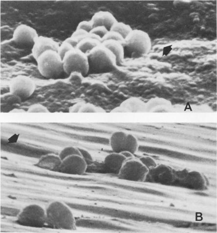
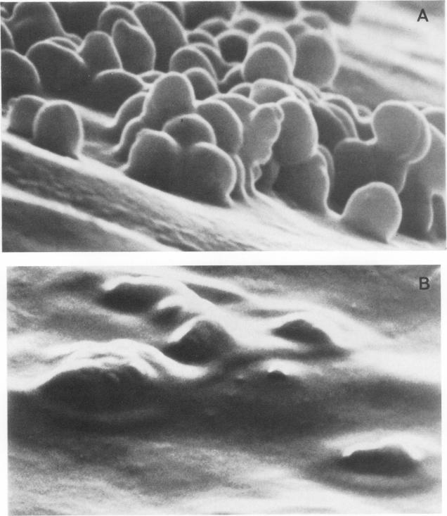
Scanning electron microscopy was used to assess the morphological features of coagulase-negative staphylococci adherent to polyvinylchloride intravascular catheter specimens. Clinical specimens were obtained by using patient catheters from which coagulase-negative staphylococci (greater than or equal to 15 colonies per catheter) grew on semiquantitative blood agar roll cultures. In vitro specimens were prepared by a previously published technique in which sterile polyvinylchloride catheters were immersed in 10(6) CFU of coagulase-negative staphylococci per ml suspended in phosphate-buffered saline. Unused sterile polyvinylchloride catheters were also examined. Scanning electron microscopy of unused sterile polyvinylchloride catheters demonstrated multiple linear surface irregularities. Scanning electron microscopy of infected patient catheters showed a diffuse amorphous material covering the entire surface and the presence of bacteria which appeared anchored to that surface by several different means. These included a slime layer, "foot" processes, and lodgement in surface irregularities. Scanning electron microscopy of in vitro specimens demonstrated no background surface coating, but it did show attachment of cocci to the surface by the same mechanisms as described for clinical specimens. These observations of similar means of attachment in clinical and in vitro specimens suggest that intrinsic catheter surface properties, bacterial surface features, and perhaps coating with host substances may all play a role in bacterial attachment to intravascular catheters. More sophisticated analysis of these interactions may clarify mechanisms of pathogenesis.
采用扫描电子显微镜评估凝固酶阴性葡萄球菌附着于聚氯乙烯血管内导管标本的形态学特征。临床标本通过使用患者的导管获取,这些导管在半定量血琼脂滚动培养中培养出凝固酶阴性葡萄球菌(每个导管菌落数大于或等于15个)。体外标本通过先前发表的技术制备,即将无菌聚氯乙烯导管浸入每毫升含10(6) CFU凝固酶阴性葡萄球菌的磷酸盐缓冲盐水中。未使用的无菌聚氯乙烯导管也进行了检查。未使用的无菌聚氯乙烯导管的扫描电子显微镜显示出多个线性表面不规则。感染患者导管的扫描电子显微镜显示整个表面覆盖有弥漫性无定形物质,并且存在通过几种不同方式附着于该表面的细菌。这些方式包括黏液层、“足”状突起以及嵌入表面不规则处。体外标本的扫描电子显微镜显示没有背景表面涂层,但确实显示出球菌通过与临床标本相同的机制附着于表面。临床和体外标本中相似附着方式的这些观察结果表明,导管的固有表面特性、细菌表面特征以及可能的宿主物质包被可能都在细菌附着于血管内导管中起作用。对这些相互作用进行更深入的分析可能会阐明发病机制。