George J N, Lyons R M, Morgan R K
J Clin Invest. 1980 Jul;66(1):1-9. doi: 10.1172/JCI109821.
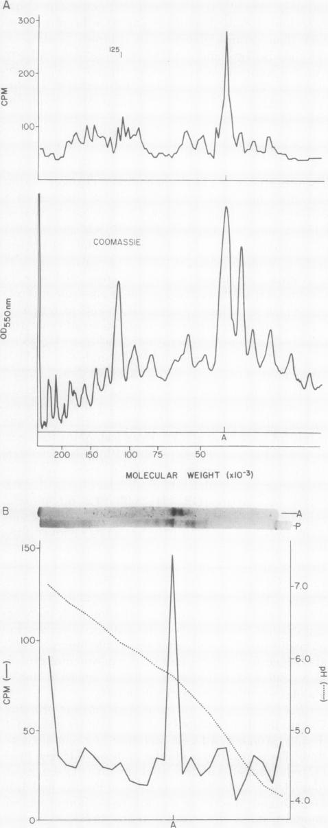
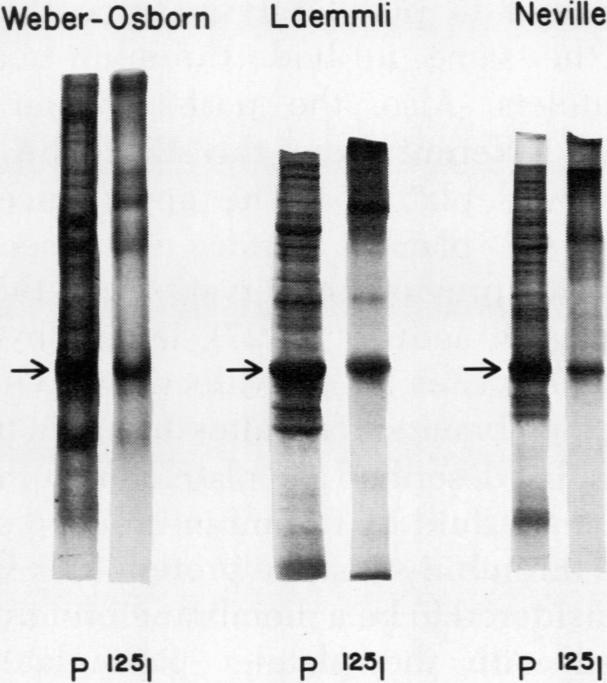
The effect of aggregation and secretion on membrane proteins was studied in washed human platelets. Reversible aggregation without secretion was stimulated by ADP and secretion without aggregation was stimulated by thrombin in the presence of EDTA. No loss of platelet surface glycoproteins occurred during reversible ADP-induced platelet aggregation, as measured by quantitative polyacrylamide gel electrophoresis analysis of platelets that were labeled with (125)I-diazotized diiodosulfanilic acid (DD(125)ISA) before ADP stimulation. Also, no new proteins became exposed on the platelet surface after ADP aggregation, as determined by DD(125)ISA labeling after stimulation. Thrombin-induced platelet secretion also caused no loss of platelet surface glycoproteins. However, after platelet secretion two new proteins were labeled by DD(125)ISA: (a) actin and (b) the 149,000-mol wt glycoprotein (termed GP-G), which is contained in platelet granules and secreted in response to thrombin. The identity of DD(125)ISA-labeled actin was confirmed by four criteria: (a) comigration with actin in three different sodium dodecyl sulfate-polyacrylamide gel electrophoresis systems, (b) elution from a particulate fraction in low ionic strength buffer, (c) co-migration with actin in isoelectric focusing, and (d) binding to DNase I. The identity of actin and its appearance on the platelet surface after thrombin-induced secretion was also demonstrated by the greater binding of an anti-actin antibody to thrombin-treated platelets, measured with (125)I-staphylococcal protein A.Therefore, major platelet membrane changes occur after secretion but not after reversible aggregation. The platelet surface changes occurring with secretion may be important in the formation of irreversible platelet aggregates and in the final retraction of the blood clot.
在洗涤过的人血小板中研究了聚集和分泌对膜蛋白的影响。ADP刺激可引起无分泌的可逆聚集,在EDTA存在的情况下,凝血酶可刺激无聚集的分泌。通过对ADP刺激前用(125)I-重氮化二碘磺胺酸(DD(125)ISA)标记的血小板进行定量聚丙烯酰胺凝胶电泳分析,发现在可逆的ADP诱导的血小板聚集中,血小板表面糖蛋白没有损失。同样,通过刺激后的DD(125)ISA标记确定,ADP聚集后血小板表面也没有新的蛋白质暴露。凝血酶诱导的血小板分泌也不会导致血小板表面糖蛋白的损失。然而,血小板分泌后,两种新的蛋白质被DD(125)ISA标记:(a)肌动蛋白和(b)149,000摩尔质量的糖蛋白(称为GP-G),它存在于血小板颗粒中,并在凝血酶作用下分泌。通过四个标准证实了DD(125)ISA标记的肌动蛋白的身份:(a)在三种不同的十二烷基硫酸钠-聚丙烯酰胺凝胶电泳系统中与肌动蛋白共迁移,(b)在低离子强度缓冲液中从颗粒部分洗脱,(c)在等电聚焦中与肌动蛋白共迁移,以及(d)与DNase I结合。用(125)I-葡萄球菌蛋白A测量,抗肌动蛋白抗体与凝血酶处理的血小板的结合增加,也证明了肌动蛋白的身份及其在凝血酶诱导的分泌后在血小板表面的出现。因此,主要的血小板膜变化发生在分泌后,而不是可逆聚集后。分泌时发生的血小板表面变化可能在不可逆血小板聚集体的形成和血凝块的最终收缩中起重要作用。