Craik C S, Buchman S R, Beychok S
Proc Natl Acad Sci U S A. 1980 Mar;77(3):1384-8. doi: 10.1073/pnas.77.3.1384.
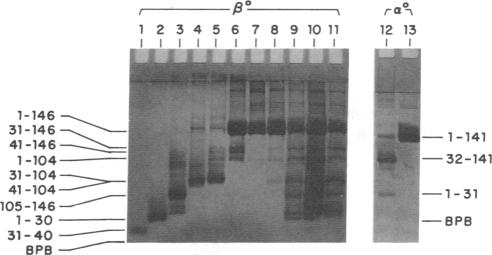
We have prepared and isolated the peptide fragments coded for by the three exons of the human beta-globin gene, using the arginine-specific protease clostripain (EC 3.4.22.8). The region encoded by the central exon (amino acid residues 31-104) contains an arginine at position 40. This site was less susceptible to cleavage than the two sites that correspond to the exon-intron boundaries, and the isolated central fragment was an approximately equimolar mixture of the entire central fragment, beta(o) (31-104), and the somewhat smaller fragment contained within it, beta(o) (41-104). This central fragment mixture bound heme stoichiometrically and tightly at micromolar concentrations, generating a strong Soret absorption band as well as a characteristic absorption band in the visible spectrum. The Soret band occurred at the same wavelength and had the same shape as in hemoglobin, exhibiting an intensity greater than (2/3) that achieved when native intact beta globin is reconstituted with heme. Nearly the full intensity was regained when an equivalent of heme was added to the unfractionated digest, suggesting that the noncovalently associated side fragments add precision to the fit of the heme pocket. Three controls were used in establishing the specificity of heme binding to the central fragment mixture. Similar, but preliminary, experiments have also been undertaken with alpha globin. A clostripain digest containing alpha(o) (1-31) and alpha(o) (32-141) bound heme, yielding a Soret band identical to that observed in alpha subunits reconstituted from the native globin chains and heme. Measurements of circular dichroism spectra as indices of secondary structure suggested a role for the side exon products in the acquisition of the native three-dimensional structure of hemoglobin. These experiments confirm a prediction of W. Gilbert that the product of the central exon of the globin gene is a complete functional domain that binds heme tightly and specifically.
我们使用精氨酸特异性蛋白酶梭菌蛋白酶(EC 3.4.22.8)制备并分离了人类β-珠蛋白基因三个外显子编码的肽片段。中央外显子编码的区域(氨基酸残基31 - 104)在第40位含有一个精氨酸。该位点比对应于外显子-内含子边界的另外两个位点更不易被切割,分离得到的中央片段是整个中央片段β(o)(31 - 104)与其中包含的稍小片段β(o)(41 - 104)的近似等摩尔混合物。这种中央片段混合物在微摩尔浓度下能化学计量且紧密地结合血红素,产生一个强的Soret吸收带以及可见光谱中的特征吸收带。Soret带出现的波长和形状与血红蛋白中的相同,其强度大于天然完整β珠蛋白与血红素重构时所达到强度的(2/3)。当向未分级的消化产物中加入等量的血红素时,几乎恢复了全部强度,这表明非共价结合的侧翼片段增加了血红素口袋适配的精确性。在确定血红素与中央片段混合物结合的特异性时使用了三个对照。也对α珠蛋白进行了类似但初步的实验。含有α(o)(1 - 31)和α(o)(32 - 141)的梭菌蛋白酶消化产物结合血红素,产生的Soret带与从天然珠蛋白链和血红素重构的α亚基中观察到的相同。作为二级结构指标的圆二色光谱测量表明,侧翼外显子产物在血红蛋白天然三维结构的形成中起作用。这些实验证实了W.吉尔伯特的一个预测,即珠蛋白基因中央外显子的产物是一个完整的功能结构域,能紧密且特异性地结合血红素。