Chao Y S, Jones A L, Hradek G T, Windler E E, Havel R J
Proc Natl Acad Sci U S A. 1981 Jan;78(1):597-601. doi: 10.1073/pnas.78.1.597.
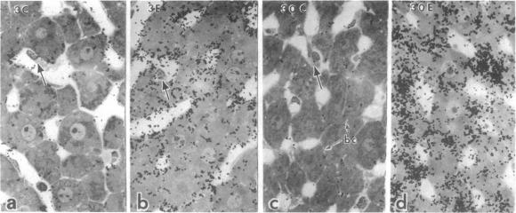
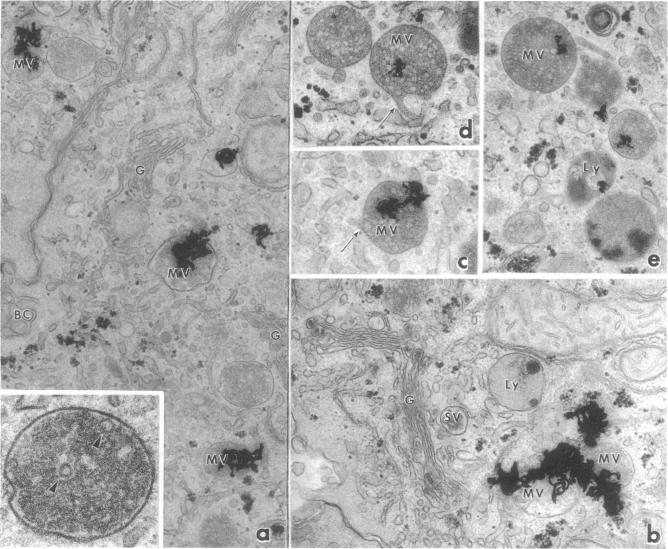
The hepatic uptake and catabolism of low density lipoproteins are stimulated severalfold in rats treated with large amounts of 17alpha-ethinylestradiol. To determine the sites within the liver at which these processes occur, (125)I-labeled human low density lipoproteins were injected intravenously into intact control and estradiol-treated rats or added to perfusates of their isolated livers. The livers were fixed by perfusion and processed for light and electron microscopic autoradiography. Distribution of autoradiographic silver grains was estimated qualitatively in light micrographs and quantitatively in electron micrographs. Many more silver grains were seen in livers from estradiol-treated than from control rats, but the processing of labeled low density lipoprotein was indistinguishable. Three minutes after intravenous injection or perfusion of livers, the grains were concentrated over the microvillous surface of parenchymal cells bordering the space of Disse. Many of these grains were within two half-distances from endocytic pits. Only 5-15% of the grains were seen over endothelial and Kupffer cells. Silver grains were also observed over vesicles beneath the plasma membrane whose size and shape suggested that they were derived from fusion of endocytic vesicles. By 15 min, grains were predominantly located in structures like multivesicular bodies in the region of the GERL (Golgi complex-endoplasmic reticulum-lysosomes) near the bile canaliculi. These bodies were packed with small vesicle-like structures and a few larger vesicles, the latter possessing a unit membrane. Between 15 and 30 min, when proteolysis of low density lipoproteins is known to begin, the initially clear matrix of the multivesicular body-like structures became dark and the structures frequently had a dense tail-like appendage. At the same time, silver grains began to appear over secondary lysosomes. These and other results indicate that the hepatic uptake of low density lipoproteins that is stimulated in rats given large amounts of estradiol follows a pathway that closely resembles that of the well-defined "LDL receptor" in cultured cells. In the liver these lipoproteins appear to be transported in endocytic vesicles; the vesicles fuse to form multivesicular body-like structures that acquire lysosomal enzymes and are converted to secondary lysosomes as the lipoproteins are degraded.
在大量服用17α-乙炔雌二醇的大鼠中,肝脏对低密度脂蛋白的摄取和分解代谢被刺激了数倍。为了确定肝脏内这些过程发生的部位,将用(125)I标记的人低密度脂蛋白静脉注射到完整的对照大鼠和经雌二醇处理的大鼠体内,或将其添加到离体肝脏的灌注液中。通过灌注固定肝脏,并进行光镜和电镜放射自显影处理。在光镜照片中定性估计放射自显影银粒的分布,在电镜照片中进行定量估计。与对照大鼠的肝脏相比,在经雌二醇处理的大鼠肝脏中可见到更多的银粒,但标记的低密度脂蛋白的处理过程并无差异。在静脉注射或灌注肝脏三分钟后,银粒集中在与狄氏间隙相邻的实质细胞的微绒毛表面。这些银粒中有许多位于距内吞小窝两个半距离之内。在内皮细胞和枯否细胞上仅可见5%-15%的银粒。在质膜下方的囊泡上也观察到银粒,其大小和形状表明它们是由内吞囊泡融合而来。到15分钟时,银粒主要位于胆小管附近的GERL(高尔基体-内质网-溶酶体)区域内的多囊体样结构中。这些结构中充满了小泡样结构和一些较大的囊泡,后者具有单位膜。在15到30分钟之间,已知低密度脂蛋白开始发生蛋白水解时,多囊体样结构最初清晰的基质变得黑暗,并且这些结构经常有一个致密的尾状附属物。与此同时,银粒开始出现在次级溶酶体上。这些以及其他结果表明,在给予大量雌二醇的大鼠中被刺激的肝脏对低密度脂蛋白的摄取遵循一条与培养细胞中明确的“低密度脂蛋白受体”途径非常相似的途径。在肝脏中,这些脂蛋白似乎在内吞囊泡中运输;囊泡融合形成多囊体样结构,这些结构获得溶酶体酶,并在脂蛋白降解时转化为次级溶酶体。