• 文献检索
  • 文档翻译
  • 深度研究
  • 学术资讯
  • Suppr Zotero 插件Zotero 插件
  • 邀请有礼
  • 套餐&价格
  • 历史记录
应用&插件
Suppr Zotero 插件Zotero 插件浏览器插件Mac 客户端Windows 客户端微信小程序
定价
高级版会员购买积分包购买API积分包
服务
文献检索文档翻译深度研究API 文档MCP 服务
关于我们
关于 Suppr公司介绍联系我们用户协议隐私条款
关注我们

Suppr 超能文献

核心技术专利:CN118964589B侵权必究
粤ICP备2023148730 号-1Suppr @ 2026

文献检索

告别复杂PubMed语法,用中文像聊天一样搜索,搜遍4000万医学文献。AI智能推荐,让科研检索更轻松。

立即免费搜索

文件翻译

保留排版,准确专业,支持PDF/Word/PPT等文件格式,支持 12+语言互译。

免费翻译文档

深度研究

AI帮你快速写综述,25分钟生成高质量综述,智能提取关键信息,辅助科研写作。

立即免费体验

计算机增强视频显微镜:可以实时生成数字处理的显微镜图像。

Computer-enhanced video microscopy: digitally processed microscope images can be produced in real time.

作者信息

Walter R J, Berns M W

出版信息

Proc Natl Acad Sci U S A. 1981 Nov;78(11):6927-31. doi: 10.1073/pnas.78.11.6927.

DOI:10.1073/pnas.78.11.6927
PMID:6947267
原文链接:https://pmc.ncbi.nlm.nih.gov/articles/PMC349165/
Abstract

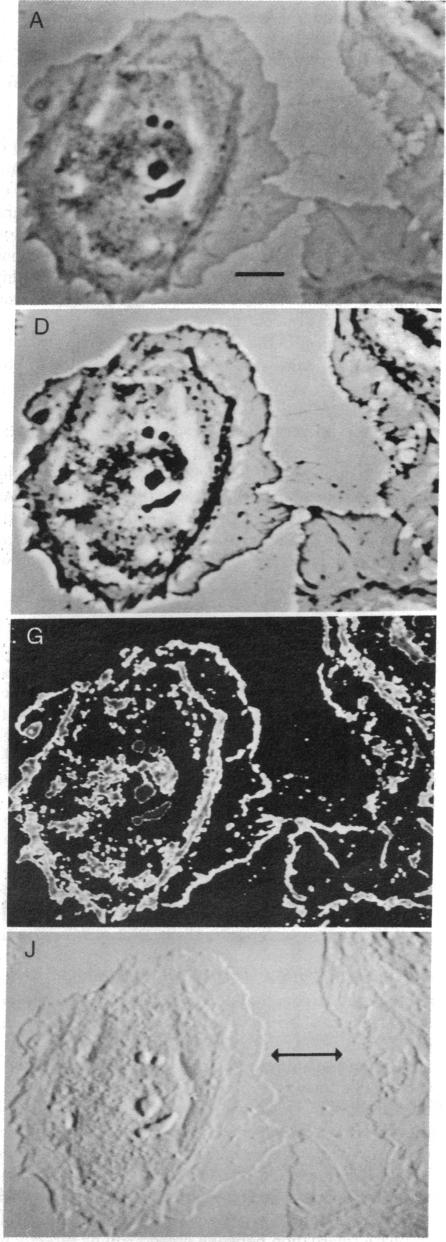
Digital processing techniques can be used to greatly enhance the available information in an optical image. Although this technology has been routinely used in many fields for a number of years, little application of digital image-processing techniques have been made toward analysis and enhancement of the types of images seen most often by the research biologist. We describe here a computer-based video microscope system that is capable of performing extensive manipulation and enhancement of microscope images in real time. The types of manipulations possible with these techniques greatly surpass the enhancement capabilities of photographic or video techniques alone. The speed and flexibility of this system enables experimental manipulation of the microscopic specimen based on its live processed image. These features greatly extend the power and versatility of the light microscope.

摘要

数字处理技术可用于大幅增强光学图像中的可用信息。尽管这项技术多年来已在许多领域得到常规应用,但数字图像处理技术在分析和增强研究生物学家最常看到的图像类型方面的应用却很少。我们在此描述一种基于计算机的视频显微镜系统,它能够实时对显微镜图像进行广泛的处理和增强。这些技术所能实现的处理类型大大超越了仅靠摄影或视频技术的增强能力。该系统的速度和灵活性使得能够根据实时处理后的图像对微观标本进行实验操作。这些特性极大地扩展了光学显微镜的功能和通用性。

https://cdn.ncbi.nlm.nih.gov/pmc/blobs/9b76/349165/2f96a552e423/pnas00662-0391-b.jpg
https://cdn.ncbi.nlm.nih.gov/pmc/blobs/9b76/349165/da777e6cb0e0/pnas00662-0391-a.jpg
https://cdn.ncbi.nlm.nih.gov/pmc/blobs/9b76/349165/2f96a552e423/pnas00662-0391-b.jpg
https://cdn.ncbi.nlm.nih.gov/pmc/blobs/9b76/349165/da777e6cb0e0/pnas00662-0391-a.jpg
https://cdn.ncbi.nlm.nih.gov/pmc/blobs/9b76/349165/2f96a552e423/pnas00662-0391-b.jpg

相似文献

1
Computer-enhanced video microscopy: digitally processed microscope images can be produced in real time.计算机增强视频显微镜:可以实时生成数字处理的显微镜图像。
Proc Natl Acad Sci U S A. 1981 Nov;78(11):6927-31. doi: 10.1073/pnas.78.11.6927.
2
Robert Feulgen Prize Lecture 1995. Electronic light microscopy: present capabilities and future prospects.1995年罗伯特·福尔根奖讲座。电子光学显微镜:当前的能力与未来的前景。
Histochem Cell Biol. 1995 Aug;104(2):97-137. doi: 10.1007/BF01451571.
3
A time-lapse, image digitization videomicroscope system based on a mini computer with large peripheral memory.一种基于带有大容量外部存储器的微型计算机的延时图像数字化视频显微镜系统。
Comput Biol Med. 1985;15(4):177-85. doi: 10.1016/0010-4825(85)90059-9.
4
A flexible, computer-controlled video microscope capable of quantitative spatial, temporal, and spectral measurements.一种灵活的、计算机控制的视频显微镜,能够进行定量的空间、时间和光谱测量。
Clin Chem. 1981 Sep;27(9):1558-68.
5
Digital light microscopy: prerequisite for optimum contrast enhancement and increase of resolution.数字光学显微镜:实现最佳对比度增强和分辨率提高的先决条件。
Exp Gerontol. 2001 Jul;36(7):1199-217. doi: 10.1016/s0531-5565(01)00087-0.
6
Video-enhanced light microscopy and its applications in cell biology.视频增强光学显微镜及其在细胞生物学中的应用。
J Cell Sci. 1988 Feb;89 ( Pt 2):129-50. doi: 10.1242/jcs.89.2.129.
7
An automated microscope for quantitative cytology combining television image analysis and stage scanning microphotometry.
J Histochem Cytochem. 1979 Jan;27(1):136-43. doi: 10.1177/27.1.374568.
8
Video microscopy, video cameras, and image enhancement.视频显微镜、摄像机和图像增强。
Cold Spring Harb Protoc. 2014 Apr 1;2014(4):359-71. doi: 10.1101/pdb.top081448.
9
Video microscopy of living cells and dynamic molecular assemblies.活细胞和动态分子组装体的视频显微镜观察。
Appl Opt. 1987 Aug 15;26(16):3219-25. doi: 10.1364/AO.26.003219.
10
Video microscopic image processing facilitates the evaluation of light microscopic autoradiography at high magnification.视频显微图像处理有助于在高放大倍数下对光学显微镜放射自显影片进行评估。
J Microsc. 1988 Jun;150(Pt 3):245-9. doi: 10.1111/j.1365-2818.1988.tb04643.x.

引用本文的文献

1
Video microscopy: an old story with a bright biological future.视频显微镜:一段有着光明生物前景的古老故事。
Biomed Eng Online. 2025 Apr 16;24(1):44. doi: 10.1186/s12938-025-01375-8.
2
Towards smart biomanufacturing: a perspective on recent developments in industrial measurement and monitoring technologies for bio-based production processes.迈向智能生物制造:生物基生产过程中工业测量和监测技术的最新发展透视。
J Ind Microbiol Biotechnol. 2020 Nov;47(11):947-964. doi: 10.1007/s10295-020-02308-1. Epub 2020 Sep 7.
3
Laser Scissors and Tweezers to Study Chromosomes: A Review.

本文引用的文献

1
Video-enhanced contrast, differential interference contrast (AVEC-DIC) microscopy: a new method capable of analyzing microtubule-related motility in the reticulopodial network of Allogromia laticollaris.视频增强对比度、微分干涉差(AVEC-DIC)显微镜检查法:一种能够分析大颈异网菌伪足网络中微管相关运动的新方法。
Cell Motil. 1981;1(3):291-302. doi: 10.1002/cm.970010303.
2
Video-enhanced contrast polarization (AVEC-POL) microscopy: a new method applied to the detection of birefringence in the motile reticulopodial network of Allogromia laticollaris.视频增强对比偏振(AVEC-POL)显微镜:一种应用于检测阔颈异网菌活动的网状伪足网络中双折射的新方法。
Cell Motil. 1981;1(3):275-89. doi: 10.1002/cm.970010302.
3
用于研究染色体的激光剪刀和镊子:综述
Front Bioeng Biotechnol. 2020 Jul 16;8:721. doi: 10.3389/fbioe.2020.00721. eCollection 2020.
4
Interactive atlas of histology: a tool for self-directed learning, practice, and self-assessment.组织学互动图谱:自我导向学习、实践和自我评估的工具。
J Chiropr Educ. 2007 Spring;21(1):12-8. doi: 10.7899/1042-5055-21.1.12.
5
Individual sarcomere length determination from isolated cardiac cells using high-resolution optical microscopy and digital image processing.使用高分辨率光学显微镜和数字图像处理技术从分离的心肌细胞中测定单个肌节长度。
Biophys J. 1982 Dec;40(3):233-44. doi: 10.1016/S0006-3495(82)84478-0.
6
Laser irradiation of centrosomes in newt eosinophils: evidence of centriole role in motility.蝾螈嗜酸性粒细胞中心体的激光照射:中心粒在运动中作用的证据
J Cell Biol. 1984 Jun;98(6):1999-2010. doi: 10.1083/jcb.98.6.1999.
7
Mitochondrial fluorescence patterns in rhodamine 6G-stained myocardial cells in vitro. Analysis by real-time computer video microscopy and laser microspot excitation.
Cell Biophys. 1984 Dec;6(4):263-77. doi: 10.1007/BF02788632.
8
The study of epithelial function by quantitative light microscopy.通过定量光学显微镜对上皮功能的研究。
Pflugers Arch. 1985;405 Suppl 1:S23-7. doi: 10.1007/BF00581775.
9
Digital imaging fluorescence microscopy: spatial heterogeneity of photobleaching rate constants in individual cells.数字成像荧光显微镜:单个细胞中光漂白速率常数的空间异质性
J Cell Biol. 1985 Apr;100(4):1309-23. doi: 10.1083/jcb.100.4.1309.
10
The distribution of [3H]kainate binding sites in primate hippocampus is similar to the distribution of both Ca2+-sensitive and Ca2+-insensitive [3H]kainate binding sites in rat hippocampus.
Neurochem Res. 1986 Jul;11(7):1073-82. doi: 10.1007/BF00965595.
Classification of cultured mammalian cells by shape analysis and pattern recognition.
通过形状分析和模式识别对培养的哺乳动物细胞进行分类。
Proc Natl Acad Sci U S A. 1980 Mar;77(3):1516-20. doi: 10.1073/pnas.77.3.1516.
4
Video image processing greatly enhances contrast, quality, and speed in polarization-based microscopy.视频图像处理极大地提高了基于偏振的显微镜检查中的对比度、质量和速度。
J Cell Biol. 1981 May;89(2):346-56. doi: 10.1083/jcb.89.2.346.
5
The zeiss-Nomarski differential interference equipment for transmitted-light microscopy.用于透射光显微镜的蔡司-诺马斯基微分干涉仪设备。
Z Wiss Mikrosk. 1969 Nov;69(4):193-221.
6
The analysis, synthesis, and description of biological images.生物图像的分析、合成与描述。
Ann N Y Acad Sci. 1966 Jan 31;128(3):984-1012. doi: 10.1111/j.1749-6632.1965.tb11712.x.
7
The analysis of cell images.细胞图像分析。
Ann N Y Acad Sci. 1966 Jan 31;128(3):1035-53. doi: 10.1111/j.1749-6632.1965.tb11715.x.
8
Quantitative analysis of micrographs by computer graphics.通过计算机图形学对显微照片进行定量分析。
J Microsc. 1975 Nov;105(2):205-13. doi: 10.1111/j.1365-2818.1975.tb04051.x.
9
Calcium ion distribution in cytoplasm visualised by aequorin: diffusion in cytosol restricted by energized sequestering.用水母发光蛋白可视化细胞质中的钙离子分布:通过活跃的螯合作用限制其在胞质溶胶中的扩散。
Science. 1975 Dec 19;190(4220):1204-6. doi: 10.1126/science.1198106.
10
Computer-assisted karyotyping.
J Reprod Med. 1976 Jul;17(1):53-7.