Progulske A, Holt S C
J Bacteriol. 1980 Aug;143(2):1003-18. doi: 10.1128/jb.143.2.1003-1018.1980.
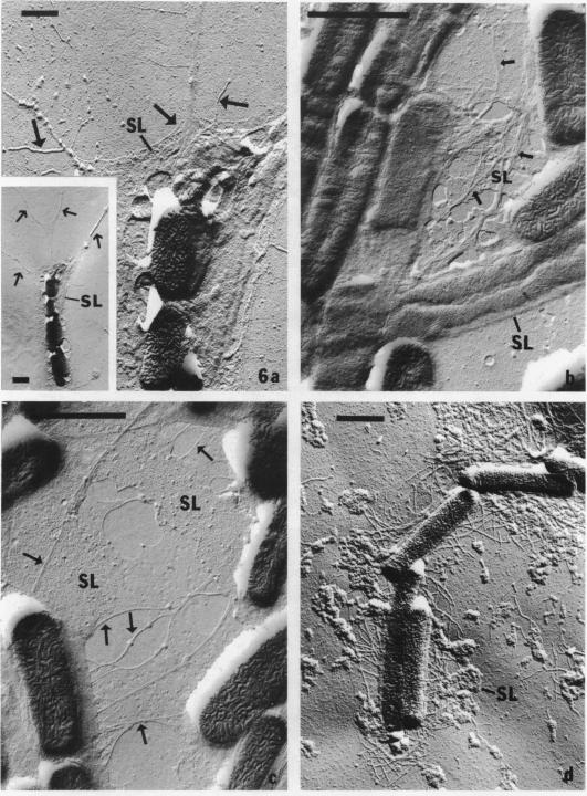
The morphology of Eikenella corrodens 333/54-55 (ATCC 23834) and two human periodontal lesion isolates, strains 470 and 373, was examined by transmission and scanning electron microscopy. All strains exhibited a cell envelope characteristic of gram-negative bacteria. Staining with ruthenium red and alcian blue revealed a loosely organized fibrous slime layer associated with the outer surface of the outer membrane. Slime "stabilization" was achieved by incubation of cells with antisera prepared against whole cells of the Eikenella strains. The stabilized slime appeared as a thick, electron-opaque layer juxtaposed to the outer membrane. Negative staining and heavy metal shadow-casting revealed an interwoven network of fibrils approximately 4 nm in diameter. These fibrils appeared to represent subunits of a larger fibril. Scanning electron microscopy after antibody slime stabilization confirmed the presence and location of the slime layer.
采用透射电子显微镜和扫描电子显微镜对腐蚀艾肯菌333/54 - 55(ATCC 23834)以及两株从人类牙周病变中分离出的菌株470和373的形态进行了检查。所有菌株均呈现革兰氏阴性菌的细胞包膜特征。用钌红和阿尔辛蓝染色显示,在外膜外表面存在一个结构松散的纤维状黏液层。通过将细胞与针对艾肯菌菌株全细胞制备的抗血清孵育来实现黏液“稳定化”。稳定后的黏液表现为与外膜相邻的一层厚的、电子不透明层。负染色和重金属投影显示出直径约4纳米的交织纤维网络。这些纤维似乎代表更大纤维的亚基。抗体黏液稳定化后的扫描电子显微镜证实了黏液层的存在和位置。