Stanley J R, Yaar M, Hawley-Nelson P, Katz S I
J Clin Invest. 1982 Aug;70(2):281-8. doi: 10.1172/jci110615.
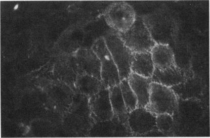
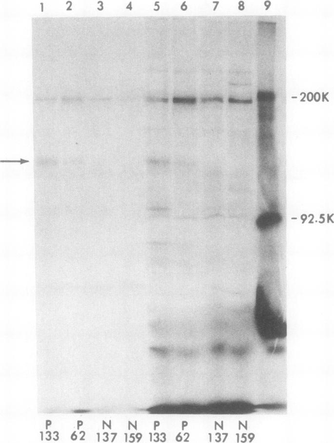
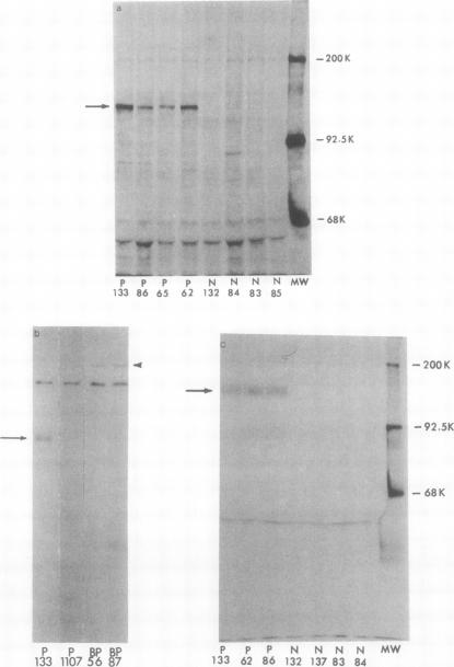
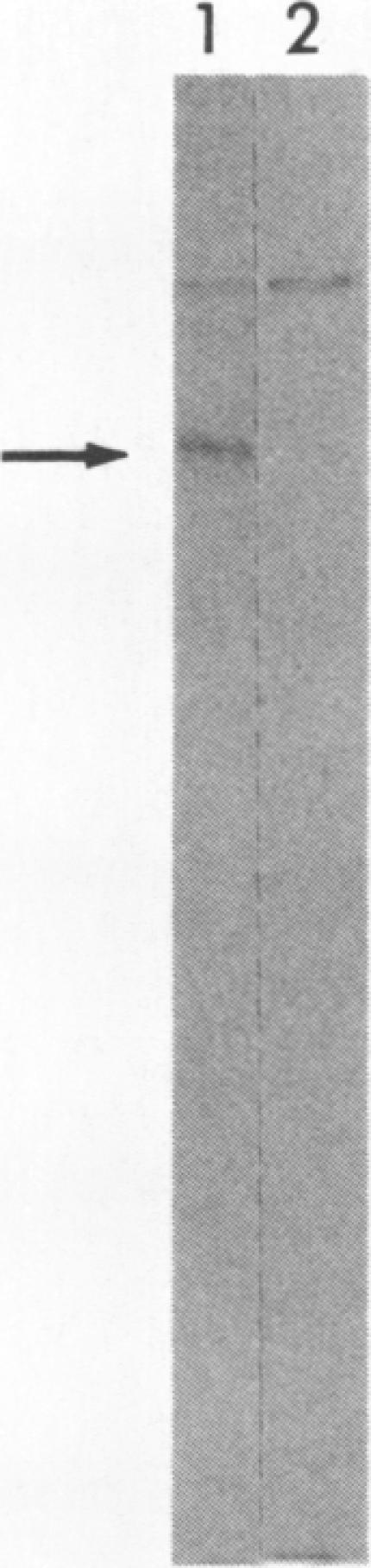
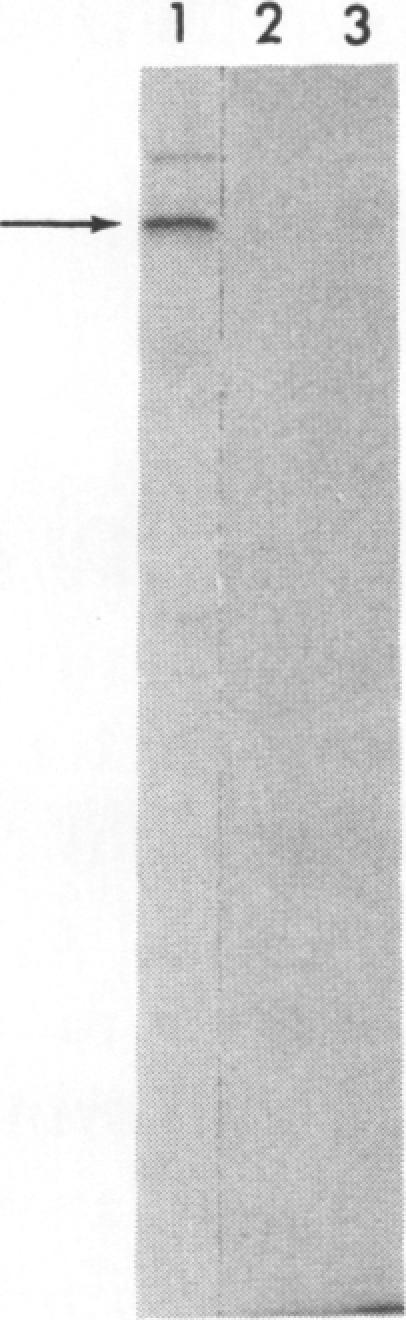
Pemphigus is an antibody-mediated autoimmune skin disease in which loss of cell-to-cell contacts in the epidermis results in blister formation. Patients with pemphigus develop antibodies that bind to the keratinocyte cell surface, the site of primary pathology. The purpose of this study was to characterize the antigen(s) to which pemphigus antibodies bind. Because we could detect pemphigus antigen by indirect immunofluorescence on the surface of multiply-passaged cells in cultures of both a spontaneously transformed mouse keratinocyte cell line (Pam) and normal human epidermal cells, we used these cells as a source of antigen. In order to demonstrate biosynthesis of antigen and to characterize the antigen(s), we radiolabeled cell cultures with [(14)C]glucosamine or d-[2-(3)H]mannose and used different pemphigus sera to immunoprecipitate antigen from nonionic detergent extracts of these labeled cells. Specifically precipitated radiolabeled molecules were identified using sodium dodecyl sulfate (SDS) polyacrylamide gel electrophoresis (PAGE) and fluorography. Sera from five of seven pemphigus patients specifically precipitated (from extracts of both Pam cells and human epidermal cells) a molecule that, when reduced, was approximately 130 kD, whereas seven normal human sera and two pemphigoid sera did not precipitate this molecule. The findings that (a) these precipitated molecules comigrated on SDS-PAGE and that (b) the 130-kD molecule could no longer be precipitated from cell extracts that had been previously reacted with a pemphigus serum, indicate that reactive pemphigus sera bind the same molecule. The molecule was not detected in the culture medium of these cells. This finding, along with the cell surface immunofluorescence pattern, suggests that the antigen is bound to the cell surface. Cultured mouse and human fibroblasts do not synthesize the antigen. The antigen contains protein because it was degraded by V8 protease and chymotrypsin, and it could also be labeled with [(14)C]amino acids. It is probably not a sulfated proteoglycan because it did not label with (35)SO(4). Taken together, these data indicate that some, but not all, pemphigus sera bind a specific cell surface glycoprotein that is synthesized by keratinocytes.
天疱疮是一种抗体介导的自身免疫性皮肤病,其中表皮中细胞间连接的丧失导致水疱形成。天疱疮患者产生与角质形成细胞表面结合的抗体,该表面是主要病理部位。本研究的目的是鉴定天疱疮抗体所结合的抗原。由于我们可以通过间接免疫荧光在自发转化的小鼠角质形成细胞系(Pam)和正常人表皮细胞培养物中多次传代的细胞表面检测到天疱疮抗原,因此我们使用这些细胞作为抗原来源。为了证明抗原的生物合成并鉴定该抗原,我们用[(14)C]葡萄糖胺或d-[2-(3)H]甘露糖对细胞培养物进行放射性标记,并使用不同的天疱疮血清从这些标记细胞的非离子去污剂提取物中免疫沉淀抗原。使用十二烷基硫酸钠(SDS)聚丙烯酰胺凝胶电泳(PAGE)和荧光自显影鉴定特异性沉淀的放射性标记分子。七名天疱疮患者中的五名患者的血清(从Pam细胞和人表皮细胞的提取物中)特异性沉淀出一种分子,该分子还原后约为130kD,而七份正常人血清和两份类天疱疮血清未沉淀出该分子。这些沉淀分子在SDS-PAGE上共迁移以及先前与天疱疮血清反应过的细胞提取物中不再能沉淀出130-kD分子的发现表明,反应性天疱疮血清结合相同的分子。在这些细胞的培养基中未检测到该分子。这一发现与细胞表面免疫荧光模式一起表明,抗原与细胞表面结合。培养的小鼠和人成纤维细胞不合成该抗原。该抗原含有蛋白质,因为它被V8蛋白酶和胰凝乳蛋白酶降解,并且它也可以用[(14)C]氨基酸标记。它可能不是硫酸化蛋白聚糖,因为它不能用(35)SO(4)标记。综上所述,这些数据表明,部分(而非全部)天疱疮血清结合一种由角质形成细胞合成的特异性细胞表面糖蛋白。