• 文献检索
  • 文档翻译
  • 深度研究
  • 学术资讯
  • Suppr Zotero 插件Zotero 插件
  • 邀请有礼
  • 套餐&价格
  • 历史记录
应用&插件
Suppr Zotero 插件Zotero 插件浏览器插件Mac 客户端Windows 客户端微信小程序
定价
高级版会员购买积分包购买API积分包
服务
文献检索文档翻译深度研究API 文档MCP 服务
关于我们
关于 Suppr公司介绍联系我们用户协议隐私条款
关注我们

Suppr 超能文献

核心技术专利:CN118964589B侵权必究
粤ICP备2023148730 号-1Suppr @ 2026

文献检索

告别复杂PubMed语法,用中文像聊天一样搜索,搜遍4000万医学文献。AI智能推荐,让科研检索更轻松。

立即免费搜索

文件翻译

保留排版,准确专业,支持PDF/Word/PPT等文件格式,支持 12+语言互译。

免费翻译文档

深度研究

AI帮你快速写综述,25分钟生成高质量综述,智能提取关键信息,辅助科研写作。

立即免费体验

鹌鹑输卵管纤毛细胞轴丝中微管蛋白、肌动蛋白和肌球蛋白的免疫细胞化学定位。

Immunocytochemical localization of tubulin, actin, and myosin in axonemes of ciliated cells from quail oviduct.

作者信息

Sandoz D, Gounon P, Karsenti E, Sauron M E

出版信息

Proc Natl Acad Sci U S A. 1982 May;79(10):3198-202. doi: 10.1073/pnas.79.10.3198.

DOI:10.1073/pnas.79.10.3198
PMID:7048302
原文链接:https://pmc.ncbi.nlm.nih.gov/articles/PMC346382/
Abstract

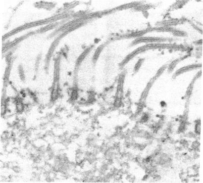
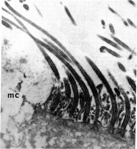
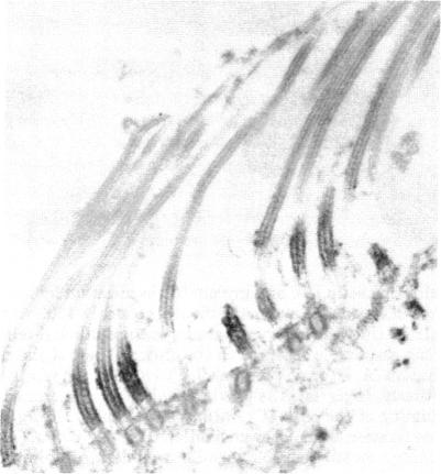
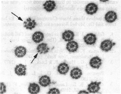
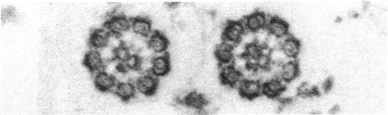
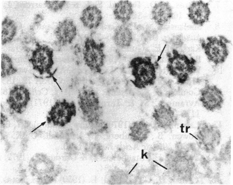
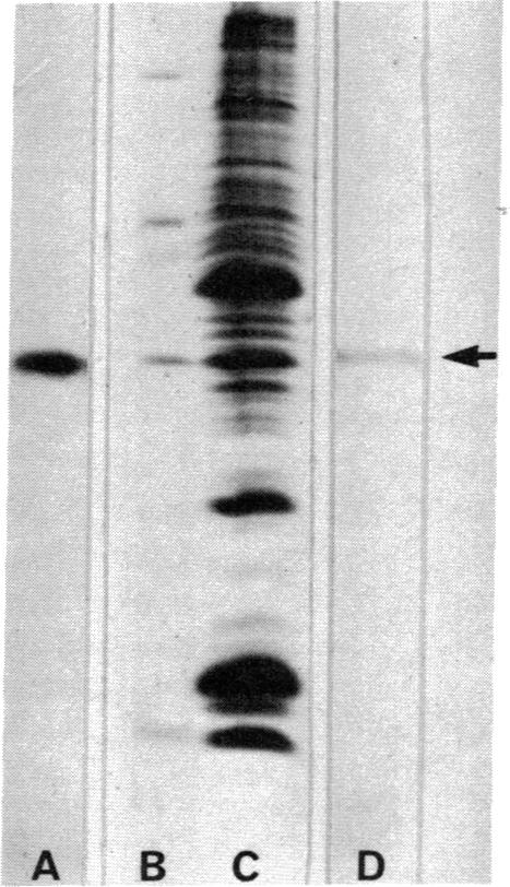
Tubulin, actin, and myosin have been localized in isolated demembranated ciliated cells from quail oviduct by immunocytochemistry in both light and electron microscopy by using purified antibodies. The peripheral doublets and the central tubules are stained by the antitubulin whereas the kinetosomes are poorly stained. Actin antibodies clearly stain the axonemes, but only on the proximal-half portion, whereas myosin antibodies stain a small area of the axonemes just above the ciliary neck region.

摘要

通过使用纯化抗体,采用免疫细胞化学方法,在光学显微镜和电子显微镜下,已将微管蛋白、肌动蛋白和肌球蛋白定位到鹌鹑输卵管分离出的去膜纤毛细胞中。抗微管蛋白可使外周双联体和中央微管染色,而基体染色较差。肌动蛋白抗体能清晰地使轴丝染色,但仅在近端一半部分,而肌球蛋白抗体则使轴丝在纤毛颈部区域上方的一小片区域染色。

https://cdn.ncbi.nlm.nih.gov/pmc/blobs/351f/346382/d847b49f4f48/pnas00449-0146-a.jpg
https://cdn.ncbi.nlm.nih.gov/pmc/blobs/351f/346382/fd1140084aed/pnas00449-0144-a.jpg
https://cdn.ncbi.nlm.nih.gov/pmc/blobs/351f/346382/2f9f7278bd33/pnas00449-0144-c.jpg
https://cdn.ncbi.nlm.nih.gov/pmc/blobs/351f/346382/83582e2e6209/pnas00449-0144-d.jpg
https://cdn.ncbi.nlm.nih.gov/pmc/blobs/351f/346382/5d4c85028a4d/pnas00449-0145-b.jpg
https://cdn.ncbi.nlm.nih.gov/pmc/blobs/351f/346382/a1a109d4a4a2/pnas00449-0145-c.jpg
https://cdn.ncbi.nlm.nih.gov/pmc/blobs/351f/346382/e4c568fd707d/pnas00449-0145-d.jpg
https://cdn.ncbi.nlm.nih.gov/pmc/blobs/351f/346382/2e06fe97680b/pnas00449-0145-e.jpg
https://cdn.ncbi.nlm.nih.gov/pmc/blobs/351f/346382/c15ed6b1a8e2/pnas00449-0145-f.jpg
https://cdn.ncbi.nlm.nih.gov/pmc/blobs/351f/346382/d847b49f4f48/pnas00449-0146-a.jpg

相似文献

1
Immunocytochemical localization of tubulin, actin, and myosin in axonemes of ciliated cells from quail oviduct.鹌鹑输卵管纤毛细胞轴丝中微管蛋白、肌动蛋白和肌球蛋白的免疫细胞化学定位。
Proc Natl Acad Sci U S A. 1982 May;79(10):3198-202. doi: 10.1073/pnas.79.10.3198.
2
Immunological detection of actin in isolated cilia from quail oviduct.鹌鹑输卵管分离纤毛中肌动蛋白的免疫检测
Biol Cell. 1986;58(1):43-52. doi: 10.1111/j.1768-322x.1986.tb00487.x.
3
Immunocytochemical localization of myosin during ciliogenesis of quail oviduct.鹌鹑输卵管纤毛发生过程中肌球蛋白的免疫细胞化学定位
Eur J Cell Biol. 1987 Jun;43(3):429-37.
4
Organization of intermediate filaments in ciliated cells from quail oviduct.鹌鹑输卵管纤毛细胞中间丝的组织
J Submicrosc Cytol. 1983 Jan;15(1):323-6.
5
Microtubule diversity in ciliated cells: evidence for its generation by post-translational modification in the axonemes of Paramecium and quail oviduct cells.纤毛细胞中的微管多样性:草履虫和鹌鹑输卵管细胞轴丝中通过翻译后修饰产生微管多样性的证据。
Biol Cell. 1991;71(1-2):227-45. doi: 10.1016/0248-4900(91)90069-y.
6
Myosin at the apical pole of ciliated epithelial cells as revealed by a monoclonal antibody.单克隆抗体揭示的纤毛上皮细胞顶端极的肌球蛋白
J Cell Biol. 1986 Aug;103(2):613-9. doi: 10.1083/jcb.103.2.613.
7
Organization of actin microfilaments in the apical border of oviduct ciliated cells.输卵管纤毛细胞顶端边界中肌动蛋白微丝的组织
Biol Cell. 1989;67(1):81-90. doi: 10.1111/j.1768-322x.1989.tb03012.x.
8
Electron microscope demonstration of tubulin in cilia and basal bodies of rat tracheal epithelium by the use of an antitubulin antibody.利用抗微管蛋白抗体对大鼠气管上皮纤毛和基体中的微管蛋白进行电子显微镜显示。
J Cell Biol. 1977 Nov;75(2 Pt 1):586-92. doi: 10.1083/jcb.75.2.586.
9
Determination of ciliary polarity precedes differentiation in the epithelial cells of quail oviduct.鹌鹑输卵管上皮细胞中纤毛极性的确定先于细胞分化。
Biol Cell. 1991;72(1-2):3-14. doi: 10.1016/0248-4900(91)90072-u.
10
Immunodetection of annexins 1 and 2 in ciliated cells from quail oviduct.鹌鹑输卵管纤毛细胞中膜联蛋白1和2的免疫检测
Biol Cell. 1992;75(1):45-54. doi: 10.1016/0248-4900(92)90123-i.

引用本文的文献

1
A Ciliary Branched Actin Network Drives Photoreceptor Disc Morphogenesis.纤毛分支肌动蛋白网络驱动光感受器盘的形态发生。
Adv Exp Med Biol. 2023;1415:507-511. doi: 10.1007/978-3-031-27681-1_74.
2
The fate of the primary cilium during myofibroblast transition.肌成纤维细胞转化过程中初级纤毛的命运。
Mol Biol Cell. 2014 Mar;25(5):643-57. doi: 10.1091/mbc.E13-07-0429. Epub 2014 Jan 8.
3
Distinct cytoskeletal domains revealed in sperm cells.精子细胞中揭示出不同的细胞骨架结构域。

本文引用的文献

1
Identification of contractile proteins in basal bodies of ciliated tracheal epithelial cells.纤毛气管上皮细胞基体中收缩蛋白的鉴定。
J Histochem Cytochem. 1980 Nov;28(11):1189-97. doi: 10.1177/28.11.7000888.
2
Involvement of contractile proteins in the changes in consistency of oocyte nucleoplasm of the newt Pleurodeles waltlii.蝾螈(Pleurodeles waltlii)卵母细胞核质稠度变化中收缩蛋白的作用。
J Cell Biol. 1981 Feb;88(2):410-21. doi: 10.1083/jcb.88.2.410.
3
The detection of DNA-binding proteins by protein blotting.通过蛋白质印迹法检测DNA结合蛋白。
J Cell Biol. 1984 Sep;99(3):1083-91. doi: 10.1083/jcb.99.3.1083.
4
Electron microscopic immunochemical localization of actin in fibroblasts in healing skin and palate wounds of beagle dog.比格犬愈合皮肤和腭部伤口中肌动蛋白在成纤维细胞内的电子显微镜免疫化学定位
Histochemistry. 1983;78(4):513-22. doi: 10.1007/BF00496203.
5
Myosin at the apical pole of ciliated epithelial cells as revealed by a monoclonal antibody.单克隆抗体揭示的纤毛上皮细胞顶端极的肌球蛋白
J Cell Biol. 1986 Aug;103(2):613-9. doi: 10.1083/jcb.103.2.613.
6
Tubulin evolution: ciliate-specific epitopes are conserved in the ciliary tubulin of Metazoa.微管蛋白的进化:纤毛虫特有的表位在后生动物的纤毛微管蛋白中保守。
J Mol Evol. 1985;22(3):220-9. doi: 10.1007/BF02099751.
7
Cytochalasin D inhibits basal body migration and ciliary elongation in quail oviduct epithelium.细胞松弛素D抑制鹌鹑输卵管上皮细胞中基体的迁移和纤毛的伸长。
Cell Tissue Res. 1990 Mar;259(3):443-54. doi: 10.1007/BF01740770.
Nucleic Acids Res. 1980 Jan 11;8(1):1-20. doi: 10.1093/nar/8.1.1.
4
Association of actin with sperm centrioles: isolation of centriolar complexes and immunofluorescent localization of actin.肌动蛋白与精子中心粒的关联:中心粒复合体的分离及肌动蛋白的免疫荧光定位
J Cell Biol. 1980 Jul;86(1):87-95. doi: 10.1083/jcb.86.1.87.
5
The early stages of absorption of injected horseradish peroxidase in the proximal tubules of mouse kidney: ultrastructural cytochemistry by a new technique.注入的辣根过氧化物酶在小鼠肾近端小管吸收的早期阶段:一种新技术的超微结构细胞化学研究
J Histochem Cytochem. 1966 Apr;14(4):291-302. doi: 10.1177/14.4.291.
6
Cleavage of structural proteins during the assembly of the head of bacteriophage T4.在噬菌体T4头部组装过程中结构蛋白的切割
Nature. 1970 Aug 15;227(5259):680-5. doi: 10.1038/227680a0.
7
Peroxidase labelled antibody and Fab conjugates with enhanced intracellular penetration.过氧化物酶标记抗体及具有增强细胞内穿透能力的Fab缀合物。
Immunochemistry. 1971 Dec;8(12):1175-9. doi: 10.1016/0019-2791(71)90395-8.
8
Microtubule assembly in the absence of added nucleotides.在未添加核苷酸的情况下微管装配。
Proc Natl Acad Sci U S A. 1973 Mar;70(3):765-8. doi: 10.1073/pnas.70.3.765.
9
Isolation of ciliated or unciliated basal bodies from the rabbit oviduct.从兔输卵管中分离纤毛或无纤毛基体。
J Cell Biol. 1974 Feb;60(2):393-404. doi: 10.1083/jcb.60.2.393.
10
A protein factor essential for microtubule assembly.微管组装所必需的一种蛋白质因子。
Proc Natl Acad Sci U S A. 1975 May;72(5):1858-62. doi: 10.1073/pnas.72.5.1858.