Bowling D, Nicholls J, Parnas I
J Physiol. 1978 Sep;282:169-80. doi: 10.1113/jphysiol.1978.sp012455.
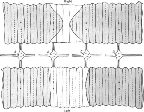
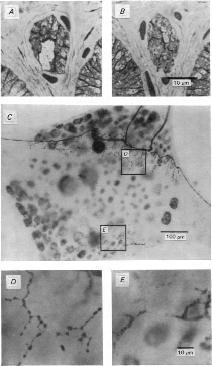
A method has been devised for killing an individual neurone in the C.N.S. of the leech by injecting it with Pronase. The technique has been used to examine the role of individual sensory and motor cells involved in producing reflex movements.1. After a neurone was injected with Pronase, either in an intact animal or an isolated ganglion, its cell body lost its resting and action potentials. Some hours later the injected cell's axons in the periphery failed to conduct impulses. In the intact animal the cell body could no longer be discerned after a few weeks.2. To test for destruction of processes within the neuropile, cells were injected first with the enzyme horseradish peroxidase (HRP) and then several hours later with Pronase. Absence of the characteristic HRP reaction product indicated that Pronase had spread throughout the arborization of the cell.3. Injection of Pronase into one cell did not produce overt electrophysiological or anatomical changes in other cells in the ganglion including neurones that were originally electrically coupled to the killed cell.4. Evidence that an individual cell was the only motoneurone supplying particular muscles was provided by destruction of that cell in otherwise intact animals, which resulted in a characteristic motor deficit in the area supplied by the killed cell. Over a period of months, functional recovery of the affected muscles occurred by way of homologous cells in adjacent ganglia.5. A further application of the technique was to trace the connexion that a particular sensory neurone makes onto two motoneurones that are electrically coupled. Normally, the sensory neurone gives rise to excitatory potentials in both post-synaptic cells. Synaptic potentials could still be recorded in one motor cell after the other had been destroyed by Pronase, indicating that synapses were made directly onto both of the motoneurones.
已设计出一种通过向水蛭中枢神经系统(C.N.S.)中的单个神经元注射链霉蛋白酶来将其杀死的方法。该技术已用于研究参与产生反射运动的单个感觉和运动细胞的作用。1. 在完整动物或分离的神经节中,向神经元注射链霉蛋白酶后,其细胞体失去静息电位和动作电位。数小时后,注射细胞在外周的轴突无法传导冲动。在完整动物中,几周后就再也无法辨认出细胞体。2. 为了测试神经纤维网内的突起是否被破坏,先向细胞注射辣根过氧化物酶(HRP),然后在数小时后注射链霉蛋白酶。没有特征性的HRP反应产物表明链霉蛋白酶已扩散到细胞的整个分支。3. 向一个细胞注射链霉蛋白酶不会在神经节中的其他细胞,包括最初与被杀死细胞电耦合的神经元中产生明显的电生理或解剖学变化。4. 在原本完整的动物中破坏单个细胞,结果在被杀死细胞所支配的区域出现特征性的运动缺陷,这证明该单个细胞是供应特定肌肉的唯一运动神经元。在几个月的时间里,受影响的肌肉通过相邻神经节中的同源细胞实现功能恢复。5. 该技术的另一个应用是追踪特定感觉神经元与两个电耦合的运动神经元之间的连接。正常情况下,感觉神经元在两个突触后细胞中都会产生兴奋性电位。在一个运动细胞被链霉蛋白酶破坏后,仍可在另一个运动细胞中记录到突触电位,这表明突触是直接在两个运动神经元上形成的。