• 文献检索
  • 文档翻译
  • 深度研究
  • 学术资讯
  • Suppr Zotero 插件Zotero 插件
  • 邀请有礼
  • 套餐&价格
  • 历史记录
应用&插件
Suppr Zotero 插件Zotero 插件浏览器插件Mac 客户端Windows 客户端微信小程序
定价
高级版会员购买积分包购买API积分包
服务
文献检索文档翻译深度研究API 文档MCP 服务
关于我们
关于 Suppr公司介绍联系我们用户协议隐私条款
关注我们

Suppr 超能文献

核心技术专利:CN118964589B侵权必究
粤ICP备2023148730 号-1Suppr @ 2026

文献检索

告别复杂PubMed语法,用中文像聊天一样搜索,搜遍4000万医学文献。AI智能推荐,让科研检索更轻松。

立即免费搜索

文件翻译

保留排版,准确专业,支持PDF/Word/PPT等文件格式,支持 12+语言互译。

免费翻译文档

深度研究

AI帮你快速写综述,25分钟生成高质量综述,智能提取关键信息,辅助科研写作。

立即免费体验

主动脉肺动脉窗的二维超声心动图诊断

Two-dimensional echocardiographic diagnosis of aorticopulmonary window.

作者信息

Satomi G, Nakamura K, Imai Y, Takao A

出版信息

Br Heart J. 1980 Mar;43(3):351-6. doi: 10.1136/hrt.43.3.351.

DOI:10.1136/hrt.43.3.351
PMID:7437182
原文链接:https://pmc.ncbi.nlm.nih.gov/articles/PMC482287/
Abstract

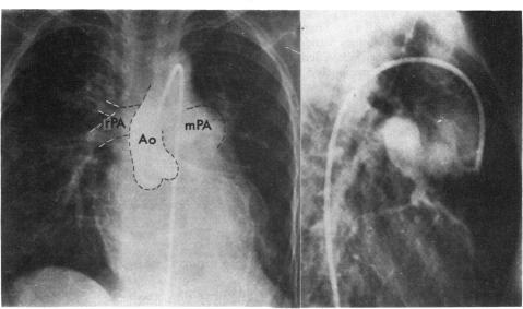
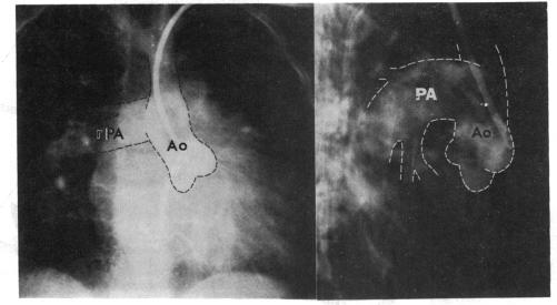
Two-dimensional echocardiography was performed in two cases of distal type aorticopulmonary window. The defect of the aorticopulmonary septum could be visualised in both cases in transverse section of the great arteries using our method. We make it a rule to study three different levels of section: plane 1, where the echo of the semilunar valve of the posterior great artery is well seen; plane 2, where the echo of the semilunar valve of the anterior great artery is clearly observed; and plane 3, where neither semilunar valve is seen. The aorticopulmonary septal defects were recognised at the level of plane 2 to plane 3 in our two cases. Two-dimensional contrast echocardiography was performed in one of the cases. The contrast entered the ascending aorta from the main pulmonary artery through the aorticopulmonary septal defect in early systole. Postoperatively, no defects were detected in the aorticopulmonary septum in either case using the same approach, and no passage of contrast into the ascending aorta from the pulmonary artery was noted in the case where contrast was injected. Accurate diagnosis of this anomaly can be made by visualisation of the defect utilising two-dimensional echocardiography. Typing of this anomaly, proximal, distal, or combined may be possible with our new approach.

摘要

对2例远端型主肺动脉窗患者进行了二维超声心动图检查。使用我们的方法,在大动脉的横切面上,2例患者均能观察到主肺动脉间隔缺损。我们通常研究三个不同的切面水平:平面1,可见后大动脉半月瓣回声;平面2,可清晰观察到前大动脉半月瓣回声;平面3,未见半月瓣回声。在我们的2例患者中,主肺动脉间隔缺损在平面2至平面3水平被识别。对其中1例患者进行了二维对比超声心动图检查。在收缩早期,造影剂通过主肺动脉间隔缺损从主肺动脉进入升主动脉。术后,采用相同方法,2例患者的主肺动脉间隔均未检测到缺损,在注射造影剂的病例中,未观察到造影剂从肺动脉进入升主动脉。利用二维超声心动图观察缺损可对该异常进行准确诊断。采用我们的新方法可能对该异常进行分型,即近端型、远端型或混合型。

https://cdn.ncbi.nlm.nih.gov/pmc/blobs/9d6c/482287/097ae1102e6a/brheartj00193-0112-b.jpg
https://cdn.ncbi.nlm.nih.gov/pmc/blobs/9d6c/482287/d4dfe49d77be/brheartj00193-0110-a.jpg
https://cdn.ncbi.nlm.nih.gov/pmc/blobs/9d6c/482287/c8e1ba74ab7d/brheartj00193-0112-a.jpg
https://cdn.ncbi.nlm.nih.gov/pmc/blobs/9d6c/482287/097ae1102e6a/brheartj00193-0112-b.jpg
https://cdn.ncbi.nlm.nih.gov/pmc/blobs/9d6c/482287/d4dfe49d77be/brheartj00193-0110-a.jpg
https://cdn.ncbi.nlm.nih.gov/pmc/blobs/9d6c/482287/c8e1ba74ab7d/brheartj00193-0112-a.jpg
https://cdn.ncbi.nlm.nih.gov/pmc/blobs/9d6c/482287/097ae1102e6a/brheartj00193-0112-b.jpg

相似文献

1
Two-dimensional echocardiographic diagnosis of aorticopulmonary window.主动脉肺动脉窗的二维超声心动图诊断
Br Heart J. 1980 Mar;43(3):351-6. doi: 10.1136/hrt.43.3.351.
2
Two-dimensional echocardiographic recognition and surgical management of aortopulmonary septal defect in the premature infant.早产儿主肺动脉间隔缺损的二维超声心动图识别与外科治疗
Thorac Cardiovasc Surg. 1982 Jun;30(3):180-3. doi: 10.1055/s-2007-1022240.
3
Distal aorticopulmonary septal defect.主动脉肺动脉间隔远端缺损
Indian Heart J. 1983 May-Jun;35(3):178-80.
4
[Echocardiographic aspects of the aortopulmonary window. Report of a case with angiocardiographic correlations].
Arch Inst Cardiol Mex. 1980 Sep-Oct;50(5):591-7.
5
Two-dimensional echocardiographic diagnosis of anomalous origin of the right pulmonary artery from the aorta: differentiation from aortopulmonary window.
J Am Coll Cardiol. 1984 Aug;4(2):351-5. doi: 10.1016/s0735-1097(84)80225-9.
6
Diagnosis of endocardial cushion defect with cross-sectional and M-mode scanning echocardiography. Differentiation from secundum atrial septal defect.经切面及M型扫描超声心动图诊断心内膜垫缺损。与继发孔型房间隔缺损的鉴别。
Br Heart J. 1976 Sep;38(9):911-20. doi: 10.1136/hrt.38.9.911.
7
Two dimensional echocardiographic assessment of communications between ascending aorta and pulmonary trunk or individual pulmonary arteries.升主动脉与肺动脉主干或各肺动脉之间交通的二维超声心动图评估。
Br Heart J. 1982 Jun;47(6):563-72. doi: 10.1136/hrt.47.6.563.
8
Aortic arch interruption with truncus arteriosus or aorticopulmonary septal defect.主动脉弓中断合并共同动脉干或主肺动脉间隔缺损。
AJR Am J Roentgenol. 1980 Nov;135(5):1011-6. doi: 10.2214/ajr.135.5.1011.
9
Interruption of the aortic arch with distal type of aorticopulmonary septal defect. A case report.
Jpn Heart J. 1980 May;21(3):443-8. doi: 10.1536/ihj.21.443.
10
Nuclear magnetic resonance imaging of the pulmonary arteries, subpulmonary region, and aorticopulmonary shunts: a comparative study with two-dimensional echocardiography and angiography.肺动脉、肺下区域及主肺动脉分流的核磁共振成像:与二维超声心动图及血管造影的对比研究
Am Heart J. 1990 May;119(5):1103-10. doi: 10.1016/s0002-8703(05)80241-8.

引用本文的文献

1
Two dimensional echocardiographic assessment of communications between ascending aorta and pulmonary trunk or individual pulmonary arteries.升主动脉与肺动脉主干或各肺动脉之间交通的二维超声心动图评估。
Br Heart J. 1982 Jun;47(6):563-72. doi: 10.1136/hrt.47.6.563.

本文引用的文献

1
Aorticopulmonary septal defect; diagnosis and report of case successfully treated.
Am J Cardiol. 1960 Feb;5:273-6. doi: 10.1016/0002-9149(60)90209-5.
2
Echocardiographic identification of aorta and main pulmonary artery in complete transposition.完全性大动脉转位中主动脉和主肺动脉的超声心动图识别
Br Heart J. 1979 Mar;41(3):356-8. doi: 10.1136/hrt.41.3.356.