• 文献检索
  • 文档翻译
  • 深度研究
  • 学术资讯
  • Suppr Zotero 插件Zotero 插件
  • 邀请有礼
  • 套餐&价格
  • 历史记录
应用&插件
Suppr Zotero 插件Zotero 插件浏览器插件Mac 客户端Windows 客户端微信小程序
定价
高级版会员购买积分包购买API积分包
服务
文献检索文档翻译深度研究API 文档MCP 服务
关于我们
关于 Suppr公司介绍联系我们用户协议隐私条款
关注我们

Suppr 超能文献

核心技术专利:CN118964589B侵权必究
粤ICP备2023148730 号-1Suppr @ 2026

文献检索

告别复杂PubMed语法,用中文像聊天一样搜索,搜遍4000万医学文献。AI智能推荐,让科研检索更轻松。

立即免费搜索

文件翻译

保留排版,准确专业,支持PDF/Word/PPT等文件格式,支持 12+语言互译。

免费翻译文档

深度研究

AI帮你快速写综述,25分钟生成高质量综述,智能提取关键信息,辅助科研写作。

立即免费体验

升主动脉与肺动脉主干或各肺动脉之间交通的二维超声心动图评估。

Two dimensional echocardiographic assessment of communications between ascending aorta and pulmonary trunk or individual pulmonary arteries.

作者信息

Smallhorn J F, Anderson R H, Macartney F J

出版信息

Br Heart J. 1982 Jun;47(6):563-72. doi: 10.1136/hrt.47.6.563.

DOI:10.1136/hrt.47.6.563
PMID:7082504
原文链接:https://pmc.ncbi.nlm.nih.gov/articles/PMC481183/
Abstract

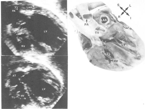
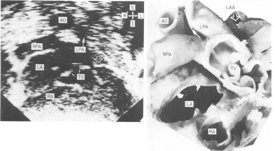
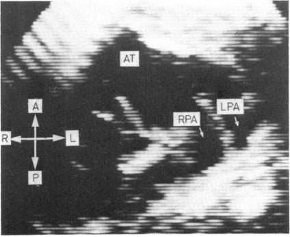
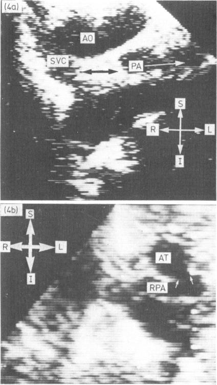
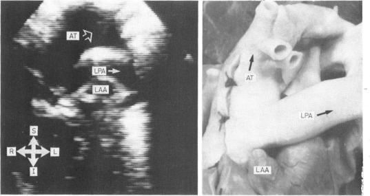
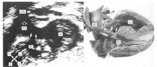
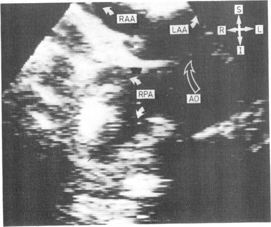
The value of two dimensional echocardiography in identifying communications between the ascending aorta and pulmonary trunk or individual pulmonary arteries was assessed in 24 children, all of whom had either angiocardiographic and surgical or angiocardiographic confirmation alone. Fourteen cases had truncus arteriosus, four aortopulmonary window, four anomalous origin of the left pulmonary artery from the ascending aorta, and two anomalous origin of the right pulmonary artery from the ascending aorta. It was possible to identify reliably each individual abnormality with a combination of suprasternal, precordial, and subcostal cuts. Problems only arose in differentiating truncus arteriosus from pulmonary atresia and ventricular septal defect when the main pulmonary artery and infundibular region of the right ventricle were extremely hypoplastic.

摘要

对24名儿童进行了评估,以确定二维超声心动图在识别升主动脉与肺动脉干或各条肺动脉之间交通方面的价值,所有这些儿童均通过心血管造影和手术或仅通过心血管造影得到确诊。14例为共同动脉干,4例为主动脉肺动脉窗,4例为左肺动脉起源于升主动脉异常,2例为右肺动脉起源于升主动脉异常。通过胸骨上、心前区和肋下切面相结合,能够可靠地识别每一种单独的异常情况。仅当主肺动脉和右心室漏斗部极度发育不全时,在鉴别共同动脉干与肺动脉闭锁及室间隔缺损方面才出现问题。

https://cdn.ncbi.nlm.nih.gov/pmc/blobs/1141/481183/d982151e6e51/brheartj00150-0059-b.jpg
https://cdn.ncbi.nlm.nih.gov/pmc/blobs/1141/481183/04334eeb1706/brheartj00150-0053-a.jpg
https://cdn.ncbi.nlm.nih.gov/pmc/blobs/1141/481183/ba5bc14534b6/brheartj00150-0054-a.jpg
https://cdn.ncbi.nlm.nih.gov/pmc/blobs/1141/481183/60d8511d7d90/brheartj00150-0054-b.jpg
https://cdn.ncbi.nlm.nih.gov/pmc/blobs/1141/481183/620b7eb88751/brheartj00150-0055-a.jpg
https://cdn.ncbi.nlm.nih.gov/pmc/blobs/1141/481183/7d0cc658d272/brheartj00150-0056-a.jpg
https://cdn.ncbi.nlm.nih.gov/pmc/blobs/1141/481183/28bde8317f6d/brheartj00150-0056-b.jpg
https://cdn.ncbi.nlm.nih.gov/pmc/blobs/1141/481183/f834216e70fc/brheartj00150-0057-a.jpg
https://cdn.ncbi.nlm.nih.gov/pmc/blobs/1141/481183/014f4b12d3fd/brheartj00150-0057-b.jpg
https://cdn.ncbi.nlm.nih.gov/pmc/blobs/1141/481183/e54abdac8213/brheartj00150-0058-a.jpg
https://cdn.ncbi.nlm.nih.gov/pmc/blobs/1141/481183/52bb4a34a89b/brheartj00150-0058-b.jpg
https://cdn.ncbi.nlm.nih.gov/pmc/blobs/1141/481183/c5f11399589c/brheartj00150-0059-a.jpg
https://cdn.ncbi.nlm.nih.gov/pmc/blobs/1141/481183/d982151e6e51/brheartj00150-0059-b.jpg

相似文献

1
Two dimensional echocardiographic assessment of communications between ascending aorta and pulmonary trunk or individual pulmonary arteries.升主动脉与肺动脉主干或各肺动脉之间交通的二维超声心动图评估。
Br Heart J. 1982 Jun;47(6):563-72. doi: 10.1136/hrt.47.6.563.
2
Two-dimensional echocardiographic prospective diagnosis of common truncus arteriosus in infants.二维超声心动图对婴儿共同动脉干的前瞻性诊断
Am J Cardiol. 1982 Dec;50(6):1380-4. doi: 10.1016/0002-9149(82)90478-7.
3
The site of origin of nonconfluent pulmonary arteries from a common arterial trunk or from the ascending aorta: its morphological significance.非融合肺动脉从共同动脉干或升主动脉发出的部位:其形态学意义。
Am J Cardiovasc Pathol. 1990;3(2):115-20.
4
The value of transthoracic echocardiography in the diagnosis of anomalous origin of the right pulmonary artery from the ascending aorta: A single center experience from China.经胸超声心动图在诊断右肺动脉起源于升主动脉异常中的价值:来自中国的单中心经验。
Int J Cardiol. 2015 Apr 1;184:750-754. doi: 10.1016/j.ijcard.2015.02.085. Epub 2015 Feb 26.
5
Anomalous origin of a pulmonary artery from the ascending aorta: surgical repair resolving pulmonary arterial hypertension.肺动脉起源于升主动脉异常:手术修复可缓解肺动脉高压
Arq Bras Cardiol. 2004 Dec;83(6):503-7; 498-502. doi: 10.1590/s0066-782x2004001800008. Epub 2005 Jan 7.
6
Two-dimensional echocardiographic study of right ventricular outflow and great artery anatomy in pulmonary atresia with ventricular septal defects and in truncus arteriosus.室间隔缺损合并肺动脉闭锁及永存动脉干患者右心室流出道和大动脉解剖结构的二维超声心动图研究
Am Heart J. 1983 Feb;105(2):281-6. doi: 10.1016/0002-8703(83)90528-8.
7
[Echocardiography findings in isolated anomalous origin of the right pulmonary artery from the ascending aorta].[升主动脉起源的孤立性右肺动脉异常的超声心动图表现]
Z Kardiol. 1991 Oct;80(10):627-9.
8
Anomalous origin of a pulmonary artery from the ascending aorta: associated anomalies and pathogenesis.肺动脉起源于升主动脉异常:相关异常及发病机制
Am J Cardiol. 1988 Apr 1;61(10):850-6. doi: 10.1016/0002-9149(88)91078-8.
9
Prenatal differentiation between truncus arteriosus (Types II and III) and pulmonary atresia with ventricular septal defect.产前区分动脉干(Ⅱ型和Ⅲ型)和室间隔缺损伴肺动脉闭锁。
Ultrasound Obstet Gynecol. 2015 Nov;46(5):564-70. doi: 10.1002/uog.14788.
10
Single origin of right and left pulmonary arteries from ascending aorta with atretic main pulmonary artery from right ventricle and left pulmonary sling.右肺动脉和左肺动脉均发自升主动脉,主肺动脉闭锁,右心室至左肺动脉间形成左肺动脉吊带。
J Cardiovasc Comput Tomogr. 2015 Nov-Dec;9(6):599-600. doi: 10.1016/j.jcct.2015.06.002. Epub 2015 Jun 30.

引用本文的文献

1
Left Pulmonary Artery from the Ascending Aorta: A Case Report and Review of Published Cases.发自升主动脉的左肺动脉:一例病例报告及已发表病例综述
J Cardiovasc Dev Dis. 2020 Dec 25;8(1):1. doi: 10.3390/jcdd8010001.
2
Intracranial arteriovenous malformation with aneurysm of the vein of Galen as cause of heart failure in infancy. Echocardiographic diagnosis and results of treatment.以大脑大静脉瘤为病因的颅内动静脉畸形导致婴儿期心力衰竭。超声心动图诊断及治疗结果。
Br Heart J. 1983 Feb;49(2):157-62. doi: 10.1136/hrt.49.2.157.
3
Cross sectional echocardiographic diagnosis of congenital heart disease in infants.

本文引用的文献

1
Two-dimensional echocardiographic diagnosis of aorticopulmonary window.主动脉肺动脉窗的二维超声心动图诊断
Br Heart J. 1980 Mar;43(3):351-6. doi: 10.1136/hrt.43.3.351.
2
Wide-angle two-dimensional echocardiographic profiles of conotruncal abnormalities.
Mayo Clin Proc. 1980 Feb;55(2):73-82.
3
Two-dimensional echocardiography in infants with persistent truncus arteriosus.永存动脉干婴儿的二维超声心动图
Br Heart J. 1981 Nov;46(5):492-7. doi: 10.1136/hrt.46.5.492.
婴儿先天性心脏病的横断面超声心动图诊断
Br Heart J. 1983 Dec;50(6):501-5. doi: 10.1136/hrt.50.6.501.
4
Diagnosis of right ventricular outflow obstruction in infants by cross sectional echocardiography.应用横断面超声心动图诊断婴儿右心室流出道梗阻
Br Heart J. 1983 Nov;50(5):416-20. doi: 10.1136/hrt.50.5.416.
5
Cross-sectional echocardiographic assessment of coarctation in the sick neonate and infant.患病新生儿和婴儿主动脉缩窄的横断面超声心动图评估。
Br Heart J. 1983 Oct;50(4):349-61. doi: 10.1136/hrt.50.4.349.
6
Aortopulmonary window, aortic origin of the right pulmonary artery, and interrupted aortic arch: detection by two-dimensional and color Doppler echocardiography in an infant.主肺动脉窗、右肺动脉的主动脉起源及主动脉弓中断:二维及彩色多普勒超声心动图在一名婴儿中的检测
Pediatr Cardiol. 1986;7(1):49-52. doi: 10.1007/BF02315481.
7
Surgical correction of anomalous origin of right pulmonary artery from aorta in a four day old neonate.一名4日龄新生儿右肺动脉起源于主动脉异常的手术矫正
Br Heart J. 1986 Jul;56(1):98-100. doi: 10.1136/hrt.56.1.98.
8
Origin of the right pulmonary artery from the ascending aorta.右肺动脉起源于升主动脉。
Ann Surg. 1987 Jul;206(1):102-13. doi: 10.1097/00000658-198707000-00016.
9
Early diagnosis and medical treatment of the persistent ductus arteriosus in infants.
Cardiovasc Intervent Radiol. 1986;9(5-6):273-8. doi: 10.1007/BF02577957.
10
Echocardiographic--angiocardiographic correlation in persistent truncus arteriosus.
Indian J Pediatr. 1989 Mar-Apr;56(2):273-6. doi: 10.1007/BF02726624.
4
Anatomical study of truncus arteriousus communis with embryological and surgical considerations.共同动脉干的解剖学研究及胚胎学与手术学考量
Br Heart J. 1976 Nov;38(11):1109-23. doi: 10.1136/hrt.38.11.1109.
5
Truncus arteriosus communis. Clinical, angiocardiographic, and pathologic findings in 100 patients.共同动脉干。100例患者的临床、心血管造影及病理表现
Am Heart J. 1976 Jul;92(1):23-38. doi: 10.1016/s0002-8703(76)80400-0.
6
Persistent truncus arteriosus. A study of 66 autopsy cases with special reference to definition and morphogenesis.
Am J Cardiol. 1977 Oct;40(4):569-78. doi: 10.1016/0002-9149(77)90073-x.
7
Cross-sectional echocardiographic diagnosis of coronary artery aneurysms in patients with the mucocutaneous lymph node syndrome.川崎病患者冠状动脉瘤的横断面超声心动图诊断
Circulation. 1979 Jan;59(1):133-9. doi: 10.1161/01.cir.59.1.133.