Dorset D L
Electron Diffraction Department, Hauptman-Woodward Medical Research Institute, Inc., Buffalo, NY 14203-1196, USA.
Proc Natl Acad Sci U S A. 1995 Oct 24;92(22):10074-8. doi: 10.1073/pnas.92.22.10074.
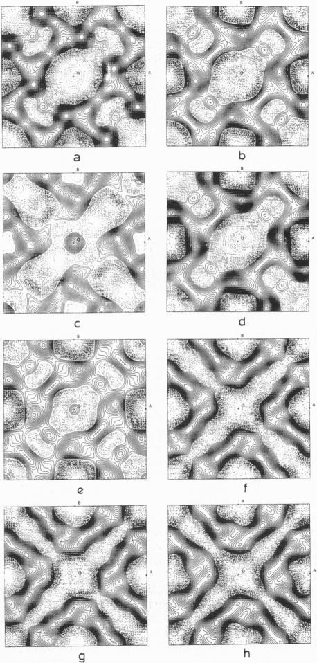
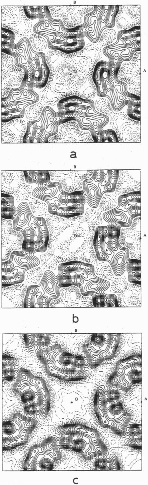
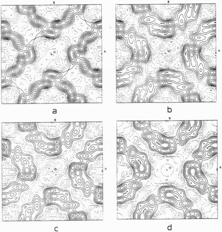
The crystal structure of halorhodopsin was determined in (centrosymmetric) projection to 6-A resolution by direct methods that use only the amplitudes of the electron diffraction pattern. A multisolution technique was used to generate initial 15-A-resolution basis sets, and after selection of the best phase set (by the closest match of magnitude of Eobs and magnitude of Ecalc), annealing of individual reflections was used to improve its accuracy. The Sayre equation was then used to expand the phase terms to 10 A, followed again by phase annealing. A final expansion with the Sayre equation enlarged this corrected phase set to 6 A. When the condition of density flatness was used to locate the best phase solution after each extension, a final structure could be observed that was quite similar to the one found earlier by analysis of electron micrographs.
通过仅使用电子衍射图案振幅的直接法,确定了嗜盐视紫红质的晶体结构在(中心对称)投影下至6埃分辨率。采用多解技术生成初始的15埃分辨率基集,在选择最佳相位集(通过观测到的E值与计算得到的E值大小的最接近匹配)后,对单个反射进行退火处理以提高其准确性。然后使用塞尔方程将相位项扩展至10埃,随后再次进行相位退火。用塞尔方程进行的最终扩展将这个校正后的相位集扩大至6埃。当在每次扩展后使用密度平坦条件来定位最佳相位解时,可以观察到最终结构与早期通过电子显微镜分析得到的结构非常相似。