• 文献检索
  • 文档翻译
  • 深度研究
  • 学术资讯
  • Suppr Zotero 插件Zotero 插件
  • 邀请有礼
  • 套餐&价格
  • 历史记录
应用&插件
Suppr Zotero 插件Zotero 插件浏览器插件Mac 客户端Windows 客户端微信小程序
定价
高级版会员购买积分包购买API积分包
服务
文献检索文档翻译深度研究API 文档MCP 服务
关于我们
关于 Suppr公司介绍联系我们用户协议隐私条款
关注我们

Suppr 超能文献

核心技术专利:CN118964589B侵权必究
粤ICP备2023148730 号-1Suppr @ 2026

文献检索

告别复杂PubMed语法,用中文像聊天一样搜索,搜遍4000万医学文献。AI智能推荐,让科研检索更轻松。

立即免费搜索

文件翻译

保留排版,准确专业,支持PDF/Word/PPT等文件格式,支持 12+语言互译。

免费翻译文档

深度研究

AI帮你快速写综述,25分钟生成高质量综述,智能提取关键信息,辅助科研写作。

立即免费体验

大鼠实验性创伤性脑损伤后凋亡性细胞死亡的证据。

Evidence of apoptotic cell death after experimental traumatic brain injury in the rat.

作者信息

Rink A, Fung K M, Trojanowski J Q, Lee V M, Neugebauer E, McIntosh T K

机构信息

Department of Neurosurgery, University of Pennsylvania, Philadelphia 19104-6316, USA.

出版信息

Am J Pathol. 1995 Dec;147(6):1575-83.

PMID:7495282
原文链接:https://pmc.ncbi.nlm.nih.gov/articles/PMC1869937/
Abstract

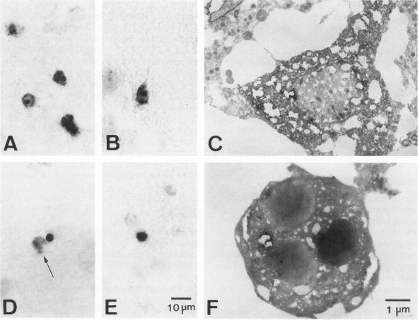
Apoptosis plays an important role in many developmental and pathological processes of the central nervous system. However, the role of apoptosis in traumatic brain injury has not been determined. Using the terminal deoxynucleotidyl transferase-mediated biotinylated deoxyuridine triphosphate nick end labeling (TUNEL) method, we detected many cells with extensive DNA fragmentation in different regions of the brains of rats subjected to experimental traumatic brain injury. Two types of TUNEL-positive cells were demonstrated by light and electron microscopy, including type I cells that displayed morphological features of necrotic cell death and type II cells that displayed morphological features of classic apoptotic cell death. TUNEL-positive cells were detectable for up to 72 hours after the initial injury. Gel electrophoresis of DNA extracted from affected areas of the injured brain containing both type I and II cells revealed only internucleosomal fragmentation at 185-bp intervals, a feature originally described in apoptotic cell death. These data suggest that apoptosis, in addition to necrotic cell death, occurs after traumatic brain injury, and that internucleosomal fragmentation of DNA may be associated with certain types of necrotic cell death.

摘要

细胞凋亡在中枢神经系统的许多发育和病理过程中发挥着重要作用。然而,细胞凋亡在创伤性脑损伤中的作用尚未明确。采用末端脱氧核苷酸转移酶介导的生物素化脱氧尿苷三磷酸缺口末端标记法(TUNEL法),我们在实验性创伤性脑损伤大鼠大脑的不同区域检测到许多具有广泛DNA片段化的细胞。通过光学显微镜和电子显微镜证实了两种TUNEL阳性细胞,包括表现出坏死性细胞死亡形态特征的I型细胞和表现出经典凋亡性细胞死亡形态特征的II型细胞。在初次损伤后长达72小时均可检测到TUNEL阳性细胞。从含有I型和II型细胞的损伤大脑受影响区域提取的DNA进行凝胶电泳显示,仅出现间隔为185 bp的核小体间片段化,这是最初在凋亡性细胞死亡中描述的特征。这些数据表明,除坏死性细胞死亡外,创伤性脑损伤后还会发生细胞凋亡,并且DNA的核小体间片段化可能与某些类型的坏死性细胞死亡有关。

https://cdn.ncbi.nlm.nih.gov/pmc/blobs/d3e2/1869937/6fa1255f3059/amjpathol00048-0066-a.jpg
https://cdn.ncbi.nlm.nih.gov/pmc/blobs/d3e2/1869937/facde41103a9/amjpathol00048-0062-a.jpg
https://cdn.ncbi.nlm.nih.gov/pmc/blobs/d3e2/1869937/b81e35889b75/amjpathol00048-0063-a.jpg
https://cdn.ncbi.nlm.nih.gov/pmc/blobs/d3e2/1869937/6fa1255f3059/amjpathol00048-0066-a.jpg
https://cdn.ncbi.nlm.nih.gov/pmc/blobs/d3e2/1869937/facde41103a9/amjpathol00048-0062-a.jpg
https://cdn.ncbi.nlm.nih.gov/pmc/blobs/d3e2/1869937/b81e35889b75/amjpathol00048-0063-a.jpg
https://cdn.ncbi.nlm.nih.gov/pmc/blobs/d3e2/1869937/6fa1255f3059/amjpathol00048-0066-a.jpg

相似文献

1
Evidence of apoptotic cell death after experimental traumatic brain injury in the rat.大鼠实验性创伤性脑损伤后凋亡性细胞死亡的证据。
Am J Pathol. 1995 Dec;147(6):1575-83.
2
Spatial and temporal profile of apoptosis following lateral fluid percussion brain injury.侧方流体冲击脑损伤后细胞凋亡的时空特征
Chin J Traumatol. 2002 Feb;5(1):24-7.
3
Specific inhibition of apoptosis after cold-induced brain injury by moderate postinjury hypothermia.损伤后适度低温对冷诱导脑损伤后细胞凋亡的特异性抑制作用。
Neurosurgery. 1998 Jul;43(1):107-14; discussion 114-5. doi: 10.1097/00006123-199807000-00070.
4
Inhibiting effect of moderate hypothermia on cell apoptosis after diffuse brain injury in rats.亚低温对大鼠弥漫性脑损伤后细胞凋亡的抑制作用
Chin J Traumatol. 2001 Feb;4(1):14-9.
5
Pharmacologic inhibition of poly(ADP-ribose) polymerase is neuroprotective following traumatic brain injury in rats.对大鼠创伤性脑损伤后,抑制聚(ADP - 核糖)聚合酶具有神经保护作用。
J Neurotrauma. 2001 Apr;18(4):369-76. doi: 10.1089/089771501750170912.
6
Evidence for apoptotic cell death in Huntington disease and excitotoxic animal models.亨廷顿病及兴奋性毒性动物模型中凋亡性细胞死亡的证据。
J Neurosci. 1995 May;15(5 Pt 2):3775-87. doi: 10.1523/JNEUROSCI.15-05-03775.1995.
7
Temporal profile of apoptotic-like changes in neurons and astrocytes following controlled cortical impact injury in the rat.大鼠控制性皮质撞击伤后神经元和星形胶质细胞凋亡样变化的时间特征。
Exp Neurol. 1999 Jul;158(1):76-88. doi: 10.1006/exnr.1999.7071.
8
Experimental brain injury induces regionally distinct apoptosis during the acute and delayed post-traumatic period.实验性脑损伤在创伤后急性期和延迟期可诱导不同区域发生凋亡。
J Neurosci. 1998 Aug 1;18(15):5663-72. doi: 10.1523/JNEUROSCI.18-15-05663.1998.
9
Differences in cell death between high and low energy brain injury in adult rats.成年大鼠高能与低能脑损伤之间细胞死亡的差异。
Acta Neurochir (Wien). 2008 Dec;150(12):1269-75;discussion 1275. doi: 10.1007/s00701-008-0147-7. Epub 2008 Nov 18.
10
Age does not influence DNA fragmentation in the hippocampus after fatal traumatic brain injury in young and aged humans compared with controls.
Clin Neuropathol. 2002 Jul-Aug;21(4):156-62.

引用本文的文献

1
Cellular and molecular mechanisms of pathological tau phosphorylation in traumatic brain injury: implications for chronic traumatic encephalopathy.创伤性脑损伤中病理性tau蛋白磷酸化的细胞和分子机制:对慢性创伤性脑病的影响
Mol Neurodegener. 2025 May 10;20(1):56. doi: 10.1186/s13024-025-00842-z.
2
Resilience of Spontaneously Hypertensive Rats to Secondary Insults After Traumatic Brain Injury: Immediate Seizures, Survival, and Stress Response.自发性高血压大鼠创伤性脑损伤后对继发性损伤的恢复力:即时癫痫发作、存活率及应激反应
Int J Mol Sci. 2025 Jan 19;26(2):829. doi: 10.3390/ijms26020829.
3
Spatial Measurement and Inhibition of Calpain Activity in Traumatic Brain Injury with an Activity-Based Nanotheranostic Platform.

本文引用的文献

1
Microscale autoradiographic method for the qualitative and quantitative analysis of apoptotic DNA fragmentation.用于凋亡DNA片段定性和定量分析的微量放射自显影方法
J Cell Physiol. 1993 Mar;154(3):519-26. doi: 10.1002/jcp.1041540310.
2
Demonstration of extensive chromatin cleavage in transplanted Morris hepatoma 7777 tissue: apoptosis or necrosis?移植的莫里斯肝癌7777组织中广泛染色质裂解的证明:凋亡还是坏死?
Am J Pathol. 1993 Mar;142(3):935-46.
3
Apoptotic photoreceptor cell death in mouse models of retinitis pigmentosa.视网膜色素变性小鼠模型中的凋亡性光感受器细胞死亡
基于活性的纳米诊疗平台对创伤性脑损伤中钙蛋白酶活性的空间测量和抑制。
ACS Nano. 2024 Sep 17;18(37):25565-25576. doi: 10.1021/acsnano.4c06052. Epub 2024 Sep 5.
4
Longitudinal assessment of mitochondrial dysfunction in acute traumatic brain injury using hyperpolarized [1- C]pyruvate.使用 1-13C 标记丙酮酸对急性创伤性脑损伤中线粒体功能障碍的纵向评估。
Magn Reson Med. 2023 Dec;90(6):2432-2442. doi: 10.1002/mrm.29794. Epub 2023 Jul 10.
5
Brain Trauma, Glucocorticoids and Neuroinflammation: Dangerous Liaisons for the Hippocampus.脑外伤、糖皮质激素与神经炎症:海马体的危险关联
Biomedicines. 2022 May 15;10(5):1139. doi: 10.3390/biomedicines10051139.
6
Role of Bevacizumab on Vascular Endothelial Growth Factor in Apolipoprotein E Deficient Mice after Traumatic Brain Injury.贝伐单抗对载脂蛋白 E 缺乏型创伤性脑损伤小鼠血管内皮生长因子的作用。
Int J Mol Sci. 2022 Apr 9;23(8):4162. doi: 10.3390/ijms23084162.
7
Peering into the Brain through the Retrosplenial Cortex to Assess Cognitive Function of the Injured Brain.通过压后皮质窥视大脑以评估损伤大脑的认知功能
Neurotrauma Rep. 2021 Dec 2;2(1):564-580. doi: 10.1089/neur.2021.0044. eCollection 2021.
8
The Role of Caspase Family in Acute Brain Injury: The Potential Therapeutic Targets in the Future.Caspase 家族在急性脑损伤中的作用:未来的潜在治疗靶点。
Curr Neuropharmacol. 2022;20(6):1194-1211. doi: 10.2174/1570159X19666211111121146.
9
Spatial Distribution of Neuropathology and Neuroinflammation Elucidate the Biomechanics of Fluid Percussion Injury.神经病理学和神经炎症的空间分布阐明了流体冲击伤的生物力学。
Neurotrauma Rep. 2021 Feb 8;2(1):59-75. doi: 10.1089/neur.2020.0046. eCollection 2021.
10
Neuroinflammation and Neuronal Loss in the Hippocampus Are Associated with Immediate Posttraumatic Seizures and Corticosterone Elevation in Rats.海马体中的神经炎症和神经元丢失与大鼠创伤后即刻癫痫发作及皮质酮升高有关。
Int J Mol Sci. 2021 May 30;22(11):5883. doi: 10.3390/ijms22115883.
Proc Natl Acad Sci U S A. 1994 Feb 1;91(3):974-8. doi: 10.1073/pnas.91.3.974.
4
Genetic control of programmed cell death in Drosophila.
Science. 1994 Apr 29;264(5159):677-83. doi: 10.1126/science.8171319.
5
Mild experimental brain injury in the rat induces cognitive deficits associated with regional neuronal loss in the hippocampus.
J Neurotrauma. 1993 Winter;10(4):405-14. doi: 10.1089/neu.1993.10.405.
6
Evidence of nuclear DNA fragmentation following hypoxia-ischemia in the infant rat brain, and transient forebrain ischemia in the adult gerbil.
Brain Pathol. 1994 Apr;4(2):115-22. doi: 10.1111/j.1750-3639.1994.tb00821.x.
7
Persistent memory dysfunction is associated with bilateral hippocampal damage following experimental brain injury.持续性记忆功能障碍与实验性脑损伤后双侧海马损伤有关。
Neurosci Lett. 1994 Feb 28;168(1-2):151-4. doi: 10.1016/0304-3940(94)90438-3.
8
Overexpression of BCL-2 in transgenic mice protects neurons from naturally occurring cell death and experimental ischemia.在转基因小鼠中,BCL-2的过表达可保护神经元免受自然发生的细胞死亡和实验性缺血的影响。
Neuron. 1994 Oct;13(4):1017-30. doi: 10.1016/0896-6273(94)90266-6.
9
Tumor cell proliferation and apoptosis in medulloblastoma.髓母细胞瘤中的肿瘤细胞增殖与凋亡
Acta Neuropathol. 1994;87(4):362-70. doi: 10.1007/BF00313605.
10
Altered gene expression in neurons during programmed cell death: identification of c-jun as necessary for neuronal apoptosis.程序性细胞死亡过程中神经元基因表达的改变:鉴定c-jun为神经元凋亡所必需。
J Cell Biol. 1994 Dec;127(6 Pt 1):1717-27. doi: 10.1083/jcb.127.6.1717.