Takeshita S, Rossow S T, Kearney M, Zheng L P, Bauters C, Bunting S, Ferrara N, Symes J F, Isner J M
Department of Medicine (Cardiology), St. Elizabeth's Medical Center, Tufts University School of Medicine, Boston, Massachusetts, USA.
Am J Pathol. 1995 Dec;147(6):1649-60.
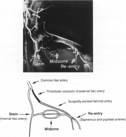
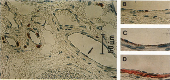
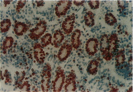
Proliferation of vascular cells has been previously shown to contribute to spontaneous development of coronary collaterals. Recent studies from several laboratories have established that collateral artery growth in both the heart and limb can be enhanced by administration of angiogenic growth factors, or therapeutic angiogenesis. In this study, we sought (1) to define the extent and time course of endothelial cell (EC) and smooth muscle cell (SMC) proliferation accompanying spontaneous collateral development during limb ischemia and (2) to determine the extent to which proliferative activity of ECs and SMCs is augmented during therapeutic angiogenesis with vascular endothelial growth factor (VEGF), a heparin-binding EC-specific mitogen. Ten days after induction of limb ischemia by surgically excising the femoral artery of rabbits, either VEGF (500 to 1000 micrograms) or saline was administered as a bolus into the iliac artery of the ischemic limb. Cellular proliferation was evaluated by bromodeoxyuridine labeling for 24 hours at day 0 (immediately before VEGF administration) and at days 3, 5, and 7 after VEGF, EC proliferation in the midzone collaterals of VEGF-treated animals increased 2.8-fold at day 5 (P < 0.05 versus control), and returned to baseline levels by day 7. SMC proliferation in midzone collaterals also increased 2.7-fold in response to VEGF (P < 0.05). No significant increase in EC or SMC proliferation was observed in either the stem or re-entry collaterals of VEGF-treated animals compared with untreated ischemic control animals. Reduction of hemodynamic deficit in the ischemic limb measured by lower limb blood pressure was documented at day 7 after VEGF (P < 0.01 versus untreated, ischemic control). These data thus (1) establish the contribution of cellular proliferation to collateral vessel development in limb ischemia and (2) support the concept that augmented cellular proliferation contributes to the enhanced formation of collateral vessels after therapeutic angiogenesis with VEGF.
血管细胞的增殖先前已被证明有助于冠状动脉侧支循环的自然形成。多个实验室最近的研究证实,通过给予血管生成生长因子或进行治疗性血管生成,可以促进心脏和肢体侧支动脉的生长。在本研究中,我们试图:(1)确定肢体缺血期间伴随侧支循环自然形成的内皮细胞(EC)和平滑肌细胞(SMC)增殖的程度和时间进程;(2)确定在用血管内皮生长因子(VEGF,一种肝素结合的EC特异性促有丝分裂原)进行治疗性血管生成期间,EC和SMC的增殖活性增强的程度。通过手术切除兔股动脉诱导肢体缺血10天后,将VEGF(500至1000微克)或生理盐水作为大剂量注入缺血肢体的髂动脉。在第0天(VEGF给药前即刻)以及VEGF给药后的第3、5和7天,通过溴脱氧尿苷标记评估细胞增殖24小时。VEGF治疗组动物中间区侧支循环中的EC增殖在第5天增加了2.8倍(与对照组相比,P<0.05),并在第7天恢复到基线水平。中间区侧支循环中的SMC增殖对VEGF的反应也增加了2.7倍(P<0.05)。与未治疗的缺血对照动物相比,VEGF治疗组动物的主干或再入侧支循环中的EC或SMC增殖均未观察到显著增加。VEGF给药后第7天记录到通过下肢血压测量的缺血肢体血流动力学缺陷的减轻(与未治疗的缺血对照相比,P<0.01)。因此,这些数据:(1)证实了细胞增殖对肢体缺血侧支血管形成的作用;(2)支持了细胞增殖增加有助于VEGF治疗性血管生成后侧支血管增强形成的概念。