• 文献检索
  • 文档翻译
  • 深度研究
  • 学术资讯
  • Suppr Zotero 插件Zotero 插件
  • 邀请有礼
  • 套餐&价格
  • 历史记录
应用&插件
Suppr Zotero 插件Zotero 插件浏览器插件Mac 客户端Windows 客户端微信小程序
定价
高级版会员购买积分包购买API积分包
服务
文献检索文档翻译深度研究API 文档MCP 服务
关于我们
关于 Suppr公司介绍联系我们用户协议隐私条款
关注我们

Suppr 超能文献

核心技术专利:CN118964589B侵权必究
粤ICP备2023148730 号-1Suppr @ 2026

文献检索

告别复杂PubMed语法,用中文像聊天一样搜索,搜遍4000万医学文献。AI智能推荐,让科研检索更轻松。

立即免费搜索

文件翻译

保留排版,准确专业,支持PDF/Word/PPT等文件格式,支持 12+语言互译。

免费翻译文档

深度研究

AI帮你快速写综述,25分钟生成高质量综述,智能提取关键信息,辅助科研写作。

立即免费体验

哈德氏腺:三百周年回顾

The harderian gland: a tercentennial review.

作者信息

Payne A P

机构信息

Department of Anatomy, Glasgow University, Scotland, UK.

出版信息

J Anat. 1994 Aug;185 ( Pt 1)(Pt 1):1-49.

PMID:7559104
原文链接:https://pmc.ncbi.nlm.nih.gov/articles/PMC1166813/
Abstract

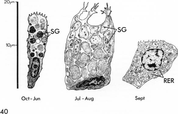
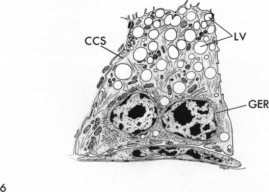
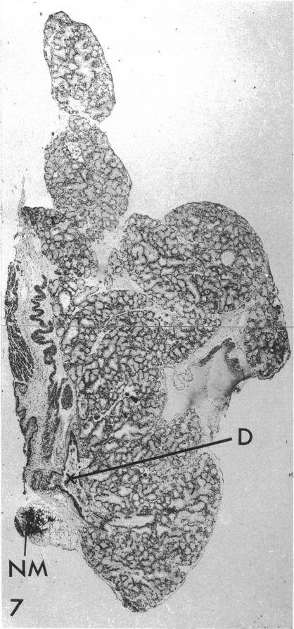
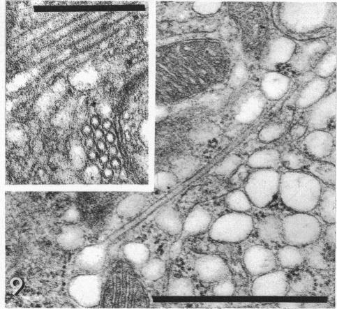
The harderian gland was first described in 1694 by Johann Jacob Harder (1656-1711). It occurs in most terrestrial vertebrates and is located within the orbit where, in some species, it is the largest structure. It may be compound tubular or compound tubuloalveolar, and its secretory duct is usually morphologically distinct only after leaving the substance of the gland to open on the surface of the nictitating membrane. The tubules of the gland are formed of a single layer of columnar epithelial cells surrounded by myoepithelial cells. The chief product(s) of the gland varies between different groups of vertebrates, and epithelial cells possess granules or vacuoles whose contents may be mucous, serous or lipid. In rodents, the gland synthesises lipids, porphyrins and indoles. In the case of lipid vacuoles, the gland is unusual in releasing these by an exocytotic mechanism. It is unclear whether the gland can act both as an exocrine and endocrine organ. There is control of gland structure and synthesis through a variety of humoral agents, including gonadal, thyroid and pituitary hormones; in addition there is a rich autonomic innervation and many neuropeptides have been identified. The proposed functions of the gland are remarkably diverse and include the gland being (1) a source of 'saliva', (2) a site of immune response, (3) a photoprotective organ, (4) part of a retinal-pineal axis, (5) a source of pheromones, (6) a source of thermoregulatory lipids, (7) a site of osmoregulation, and (8) a source of growth factors. The gland is discussed in terms of its embryology and phylogeny, and in relation to ecological variables. Several goals of future research are identified.

摘要

哈德氏腺于1694年由约翰·雅各布·哈德(1656 - 1711)首次描述。它存在于大多数陆生脊椎动物中,位于眼眶内,在某些物种中,它是眼眶内最大的结构。它可能是复管泡状腺或复管泡状腺泡状腺,其分泌导管通常仅在离开腺体实质并开口于瞬膜表面后在形态上才明显不同。腺体的小管由单层柱状上皮细胞构成,周围环绕着肌上皮细胞。腺体的主要产物在不同的脊椎动物类群中有所不同,上皮细胞含有颗粒或液泡,其内容物可能是黏液、浆液或脂质。在啮齿动物中,该腺体合成脂质、卟啉和吲哚。就脂质液泡而言,该腺体通过胞吐机制释放它们的方式很不寻常。尚不清楚该腺体是否既能作为外分泌器官又能作为内分泌器官发挥作用。通过多种体液因子,包括性腺、甲状腺和垂体激素,对腺体的结构和合成进行调控;此外,还有丰富的自主神经支配,并且已鉴定出许多神经肽。该腺体的推测功能非常多样,包括(1)“唾液”来源,(2)免疫反应场所,(3)光保护器官,(4)视网膜 - 松果体轴的一部分,(5)信息素来源,(6)体温调节脂质来源,(7)渗透调节场所,以及(8)生长因子来源。本文从其胚胎学和系统发育以及与生态变量的关系方面对该腺体进行了讨论。确定了未来研究的几个目标。

https://cdn.ncbi.nlm.nih.gov/pmc/blobs/f7d5/1166813/35cf5b1e3427/janat00135-0040-a.jpg
https://cdn.ncbi.nlm.nih.gov/pmc/blobs/f7d5/1166813/115cd9946337/janat00135-0009-a.jpg
https://cdn.ncbi.nlm.nih.gov/pmc/blobs/f7d5/1166813/8b7eec1caeb7/janat00135-0009-b.jpg
https://cdn.ncbi.nlm.nih.gov/pmc/blobs/f7d5/1166813/5f565a1a6c8e/janat00135-0010-a.jpg
https://cdn.ncbi.nlm.nih.gov/pmc/blobs/f7d5/1166813/dd1a8324b757/janat00135-0010-b.jpg
https://cdn.ncbi.nlm.nih.gov/pmc/blobs/f7d5/1166813/479cbdfb9199/janat00135-0010-c.jpg
https://cdn.ncbi.nlm.nih.gov/pmc/blobs/f7d5/1166813/a95cf1543a42/janat00135-0010-d.jpg
https://cdn.ncbi.nlm.nih.gov/pmc/blobs/f7d5/1166813/958b744adeeb/janat00135-0012-a.jpg
https://cdn.ncbi.nlm.nih.gov/pmc/blobs/f7d5/1166813/b81a6a06eba7/janat00135-0012-b.jpg
https://cdn.ncbi.nlm.nih.gov/pmc/blobs/f7d5/1166813/7a4cdde4724f/janat00135-0014-a.jpg
https://cdn.ncbi.nlm.nih.gov/pmc/blobs/f7d5/1166813/fb4e4b5f7fcb/janat00135-0014-b.jpg
https://cdn.ncbi.nlm.nih.gov/pmc/blobs/f7d5/1166813/fcfca770acc1/janat00135-0014-c.jpg
https://cdn.ncbi.nlm.nih.gov/pmc/blobs/f7d5/1166813/89ceb6ed6430/janat00135-0018-a.jpg
https://cdn.ncbi.nlm.nih.gov/pmc/blobs/f7d5/1166813/5d8e8099f795/janat00135-0019-a.jpg
https://cdn.ncbi.nlm.nih.gov/pmc/blobs/f7d5/1166813/cb122624b41e/janat00135-0020-a.jpg
https://cdn.ncbi.nlm.nih.gov/pmc/blobs/f7d5/1166813/95f742569276/janat00135-0020-b.jpg
https://cdn.ncbi.nlm.nih.gov/pmc/blobs/f7d5/1166813/bf3a344ad7dd/janat00135-0020-c.jpg
https://cdn.ncbi.nlm.nih.gov/pmc/blobs/f7d5/1166813/394182213d32/janat00135-0020-d.jpg
https://cdn.ncbi.nlm.nih.gov/pmc/blobs/f7d5/1166813/46453158ca60/janat00135-0020-e.jpg
https://cdn.ncbi.nlm.nih.gov/pmc/blobs/f7d5/1166813/bb9c453fa0a7/janat00135-0020-f.jpg
https://cdn.ncbi.nlm.nih.gov/pmc/blobs/f7d5/1166813/e626dc549b4a/janat00135-0024-a.jpg
https://cdn.ncbi.nlm.nih.gov/pmc/blobs/f7d5/1166813/907af5d70349/janat00135-0024-b.jpg
https://cdn.ncbi.nlm.nih.gov/pmc/blobs/f7d5/1166813/8234b5bd7a78/janat00135-0024-c.jpg
https://cdn.ncbi.nlm.nih.gov/pmc/blobs/f7d5/1166813/d71f9e1d9028/janat00135-0024-d.jpg
https://cdn.ncbi.nlm.nih.gov/pmc/blobs/f7d5/1166813/dcf6205acc10/janat00135-0024-e.jpg
https://cdn.ncbi.nlm.nih.gov/pmc/blobs/f7d5/1166813/5b672a2514e3/janat00135-0024-f.jpg
https://cdn.ncbi.nlm.nih.gov/pmc/blobs/f7d5/1166813/07091b86b09b/janat00135-0026-a.jpg
https://cdn.ncbi.nlm.nih.gov/pmc/blobs/f7d5/1166813/9b1387792da8/janat00135-0026-b.jpg
https://cdn.ncbi.nlm.nih.gov/pmc/blobs/f7d5/1166813/ec0fce3dbcca/janat00135-0026-c.jpg
https://cdn.ncbi.nlm.nih.gov/pmc/blobs/f7d5/1166813/6385ce9033eb/janat00135-0026-d.jpg
https://cdn.ncbi.nlm.nih.gov/pmc/blobs/f7d5/1166813/895fdf3b6ccb/janat00135-0031-a.jpg
https://cdn.ncbi.nlm.nih.gov/pmc/blobs/f7d5/1166813/daa73031133d/janat00135-0033-a.jpg
https://cdn.ncbi.nlm.nih.gov/pmc/blobs/f7d5/1166813/cb6370319df6/janat00135-0038-a.jpg
https://cdn.ncbi.nlm.nih.gov/pmc/blobs/f7d5/1166813/35cf5b1e3427/janat00135-0040-a.jpg

相似文献

1
The harderian gland: a tercentennial review.哈德氏腺:三百周年回顾
J Anat. 1994 Aug;185 ( Pt 1)(Pt 1):1-49.
2
[The mammalian Harderian gland (its structural characteristics)].[哺乳动物的哈德氏腺(其结构特征)]
Izv Akad Nauk Ser Biol. 1994 Mar-Apr(2):232-53.
3
The Harderian gland of the Cheesman's gerbil (Gerbillus cheesmani ) of the Kuwaiti desert.科威特沙漠中切斯曼沙鼠(Gerbillus cheesmani)的哈德氏腺。
Eur J Morphol. 2000 Apr;38(2):97-108. doi: 10.1076/0924-3860(200004)38:2;1-f;ft097.
4
The mammalian Harderian gland: morphology, biochemistry, function and phylogeny.哺乳动物的哈德氏腺:形态学、生物化学、功能及系统发育
Arch Histol Jpn. 1981 Sep;44(4):299-333. doi: 10.1679/aohc1950.44.299.
5
The Harderian gland: perspectives.哈德氏腺:观点
Microsc Res Tech. 1996 May 1;34(1):2-5. doi: 10.1002/(SICI)1097-0029(19960501)34:1<2::AID-JEMT2>3.0.CO;2-W.
6
The Harderian gland: Endocrine function and hormonal control.哈德氏腺:内分泌功能和激素控制。
Gen Comp Endocrinol. 2020 Oct 1;297:113548. doi: 10.1016/j.ygcen.2020.113548. Epub 2020 Jul 15.
7
The harderian gland and its excretory duct in the Wistar rat. A histological and ultrastructural study.Wistar大鼠的哈德腺及其排泄管。组织学与超微结构研究。
J Anat. 1994 Jun;184 ( Pt 3)(Pt 3):553-66.
8
Light and electron microscopic studies of the Gerbillus tarabuli (Thomas, 1902) Harderian gland.北非肥尾沙鼠(托马斯,1902年)哈德氏腺的光学显微镜和电子显微镜研究。
Zoolog Sci. 2013 Jan;30(1):53-9. doi: 10.2108/zsj.30.53.
9
Cell biology of the harderian gland.哈德氏腺的细胞生物学
Int Rev Cytol. 1996;168:1-80. doi: 10.1016/s0074-7696(08)60882-7.
10
Morphological observations on the harderian gland of the North American opossum (Didelphis virginiana).北美负鼠(弗吉尼亚负鼠)哈德氏腺的形态学观察。
Anat Embryol (Berl). 1992 Jul;186(2):145-52. doi: 10.1007/BF00174952.

引用本文的文献

1
Arterial and venous supply of the Harderian gland in hens.母鸡哈德腺的动脉和静脉供应。
Open Vet J. 2024 Jul;14(7):1553-1560. doi: 10.5455/OVJ.2024.v14.i7.4. Epub 2024 Jul 31.
2
Harderian Gland Development and Degeneration in the -Deficient Heterozygous Mouse.α-缺陷杂合小鼠的哈德氏腺发育与退化
J Dev Biol. 2024 Jun 3;12(2):16. doi: 10.3390/jdb12020016.
3
The immunoglobulin A isotype of the Arabian camel () preserves the dualistic structure of unconventional single-domain and canonical heavy chains.阿拉伯骆驼 () 的免疫球蛋白 A 同种型保留了非常规单结构域和典型重链的二元结构。

本文引用的文献

1
Quantitative studies on the effects of hormones on structure and porphyrin biosynthesis in the Harderian gland of the female golden hamster: I. The effects of ovariectomy and nitrogen administration.雌性金黄仓鼠哈德氏腺中激素对结构和卟啉生物合成影响的定量研究:I. 卵巢切除和氮给药的影响
J Anat. 1985 Oct;142:59-72.
2
THE ULTRASTRUCTURE OF THE HARDERIAN GLAND OF THE MOUSE WITH PARTICULAR REFERENCE TO THE FORMATION OF ITS SECRETORY PRODUCT.小鼠哈德氏腺的超微结构,特别涉及其分泌产物的形成
J Ultrastruct Res. 1963 Aug;49:76-98. doi: 10.1016/s0022-5320(63)80037-4.
3
On the histology and ultrastructure of the Harderian gland in rabbits.
Front Immunol. 2023 Dec 12;14:1289769. doi: 10.3389/fimmu.2023.1289769. eCollection 2023.
4
The origin of blinking in both mudskippers and tetrapods is linked to life on land.眨眼的起源与泥鰍和四足动物都与陆地生活有关。
Proc Natl Acad Sci U S A. 2023 May 2;120(18):e2220404120. doi: 10.1073/pnas.2220404120. Epub 2023 Apr 24.
5
Enzyme histochemical characterization of orbital glands in fetuses of Indian buffalo ().印度野牛胎儿眼眶腺的酶组织化学特征。
PeerJ. 2023 Apr 10;11:e15196. doi: 10.7717/peerj.15196. eCollection 2023.
6
Dimorphic frataxin and its gene regulation by sex steroids in hamsters.双态型铁调素及其在仓鼠中受性类固醇的基因调控。
Mol Genet Genomics. 2023 May;298(3):615-626. doi: 10.1007/s00438-023-02004-6. Epub 2023 Mar 16.
7
Ocular Symptoms Associated with COVID-19 Are Correlated with the Expression Profile of Mouse SARS-CoV-2 Binding Sites.与 COVID-19 相关的眼部症状与小鼠 SARS-CoV-2 结合位点的表达谱相关。
Viruses. 2023 Jan 26;15(2):354. doi: 10.3390/v15020354.
8
Apocrine secretion in the salivary glands of and other dipterans is evolutionarily conserved.蠓及其他双翅目昆虫唾液腺中的顶浆分泌在进化上是保守的。
Front Cell Dev Biol. 2023 Jan 13;10:1088055. doi: 10.3389/fcell.2022.1088055. eCollection 2022.
9
The Evanescent Bouquet of Individual Bear Fingerprint.个体熊指纹的短暂芬芳。
Animals (Basel). 2023 Jan 6;13(2):220. doi: 10.3390/ani13020220.
10
Mucosal immunology of the ocular surface.眼表的黏膜免疫学
Mucosal Immunol. 2022 Jun;15(6):1143-1157. doi: 10.1038/s41385-022-00551-6. Epub 2022 Aug 24.
家兔哈德氏腺的组织学与超微结构研究
Z Zellforsch Mikrosk Anat. 1960;52:93-104. doi: 10.1007/BF00344641.
4
The identification and electron microscopy of myoepithelial cells in the Harderian gland.哈德氏腺中肌上皮细胞的鉴定及电子显微镜观察
Anat Rec. 1958 Dec;132(4):569-83. doi: 10.1002/ar.1091320406.
5
Comparison of porphyrin producing enzyme activities in harderian glands of mice and other rodents.小鼠及其他啮齿动物哈德氏腺中卟啉生成酶活性的比较。
Proc Soc Exp Biol Med. 1958 Apr;97(4):775-8. doi: 10.3181/00379727-97-23877.
6
Pigment in the harderian glands of mice exposed to a hot environment.暴露于热环境下的小鼠哈德氏腺中的色素
Q J Exp Physiol Cogn Med Sci. 1957 Jan;42(1):24-32. doi: 10.1113/expphysiol.1957.sp001240.
7
On the mast cells in the human vascular wall: a quantitative study on changes at different ages.关于人体血管壁中的肥大细胞:不同年龄变化的定量研究。
Acta Pathol Microbiol Scand Suppl. 1955(Suppl 107):1-81.
8
Role of the thyroid and anterior pituitary glands in the maintenance of the harderian glands of the rat.甲状腺和垂体前叶在维持大鼠哈德氏腺中的作用。
Endocrinology. 1954 Nov;55(5):601-12. doi: 10.1210/endo-55-5-601.
9
The Harderian gland of the rodent Octodon degus: a structural and ultrastructural study.啮齿动物八齿鼠的哈德氏腺:一项结构和超微结构研究。
Tissue Cell. 1993;25(1):129-39. doi: 10.1016/0040-8166(93)90070-2.
10
Fibronectin synthesis in the harderian gland of the chicken.鸡哈德氏腺中纤连蛋白的合成
Poult Sci. 1993 Mar;72(3):475-82. doi: 10.3382/ps.0720475.