Nason-Burchenal K, Wolff L
Laboratory of Genetics, National Cancer Institute, National Institutes of Health, Bethesda, MD 20892.
Proc Natl Acad Sci U S A. 1993 Feb 15;90(4):1619-23. doi: 10.1073/pnas.90.4.1619.
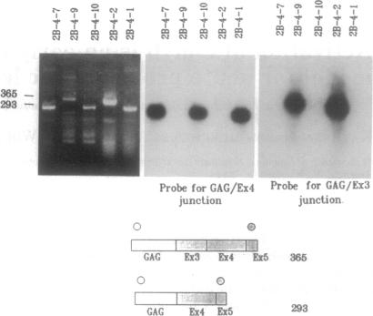
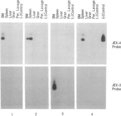
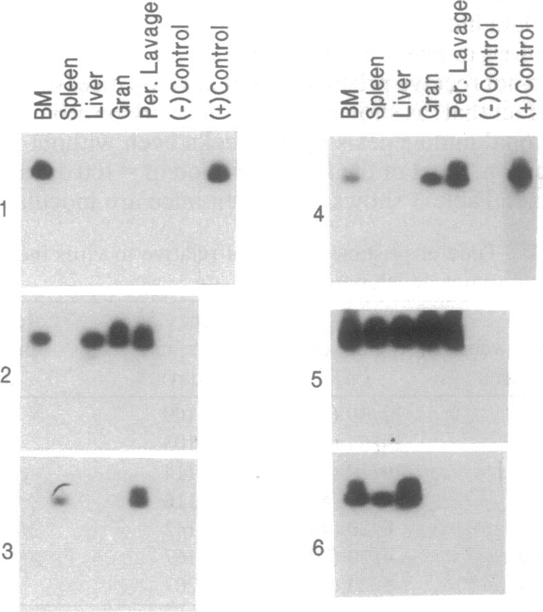
Insertional mutagenesis of c-myb by Moloney murine leukemia virus occurs in 100% of promonocytic leukemias (MMLS) induced by the virus. These leukemias, which resemble acute monocytic leukemia-M5 in humans are induced only in mice undergoing a peritoneal chronic inflammatory response. We have found that two leukemia-specific gag-myb mRNAs in MML provide molecular markers for detection of preleukemic cells in hematopoietic tissue in vivo. The two aberrant RNAs result from splicing of gag to either exon 3 or 4 of c-myb, depending on the site of proviral integration. After reverse transcription-PCR with nested primers and hybridization with specific gag-myb junction probes, one cell, having aberrant c-myb message, could be detected in a minimum of 10(5) liver cells or 10(6) spleen or bone-marrow cells. This approach was used to examine hematopoietic tissues of mice after pristane injection to induce inflammation and virus inoculation. Cells with gag-myb mRNAs could be detected as early as 2 weeks after virus inoculation. In mice receiving both pristane and virus, there was evidence of preleukemic cells in 83% of the mice by 3 weeks after virus infection. Furthermore, 100% of the mice were positive for preleukemic cells by 8 weeks, even though only 50% of mice have been shown to succumb to MML (peak time for disease latency is 12-16 weeks). Cells with these aberrant c-myb messages were initially detected in the bone marrow, but during intermediate stages of disease development these cells disseminated to the spleen, liver, and granuloma. At preleukemic times, from 3 to 8 weeks after virus infection, a lower percentage of mice were positive in the group that did not receive pristane compared with mice in the group receiving pristane. However, at 18 weeks, 100% of the mice in the group receiving virus only had evidence of cells expressing gag-myb RNA in their spleens and/or bone marrow; it is of interest that mice inoculated with virus alone never develop MML. This approach for detecting preleukemic cells will now allow the study of mechanisms by which these preleukemic cells progress to a more transformed state and, perhaps, to a more differentiated state.
莫洛尼鼠白血病病毒对c-myb的插入诱变在该病毒诱导的100%的前单核细胞白血病(MMLS)中发生。这些白血病类似于人类的急性单核细胞白血病-M5,仅在经历腹膜慢性炎症反应的小鼠中诱发。我们发现,MML中的两种白血病特异性gag-myb mRNA为体内造血组织中白血病前期细胞的检测提供了分子标记。这两种异常RNA是由gag与c-myb的外显子3或4剪接产生的,具体取决于前病毒整合的位点。用巢式引物进行逆转录PCR并与特异性gag-myb连接探针杂交后,在至少10⁵个肝细胞或10⁶个脾细胞或骨髓细胞中可检测到一个带有异常c-myb信息的细胞。该方法用于检测注射 pristane 诱导炎症并接种病毒后小鼠的造血组织。接种病毒后2周即可检测到带有gag-myb mRNA的细胞。在同时接受pristane和病毒的小鼠中,病毒感染后3周时,83%的小鼠有白血病前期细胞的证据。此外,到8周时,100%的小鼠白血病前期细胞呈阳性,尽管只有50%的小鼠已被证明死于MML(疾病潜伏期的峰值时间为12 - 16周)。带有这些异常c-myb信息的细胞最初在骨髓中被检测到,但在疾病发展的中期阶段,这些细胞扩散到脾脏、肝脏和肉芽肿。在白血病前期,即病毒感染后3至8周,未接受pristane的组中呈阳性的小鼠百分比低于接受pristane的组中的小鼠。然而,在18周时,仅接受病毒的组中100%的小鼠在其脾脏和/或骨髓中有表达gag-myb RNA的细胞的证据;有趣的是,仅接种病毒的小鼠从未发生MML。这种检测白血病前期细胞的方法现在将有助于研究这些白血病前期细胞发展为更具转化状态甚至更分化状态的机制。