Zheng Y M, Schäfer M K, Weihe E, Sheng H, Corisdeo S, Fu Z F, Koprowski H, Dietzschold B
Department of Microbiology and Immunology, Thomas Jefferson University, Philadelphia, Pennsylvania 19107-6799.
J Virol. 1993 Oct;67(10):5786-91. doi: 10.1128/JVI.67.10.5786-5791.1993.
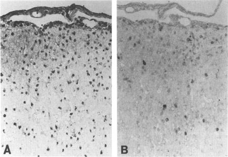
The putative role of nitric oxide in the neuropathogenesis of Borna disease was investigated by determining changes in the expression of inducible nitric oxide synthase (iNOS) mRNA and constitutively expressed NOS (cNOS) mRNA in brains of Borna disease virus (BDV)-infected rats. iNOS mRNA was not detected in normal rat brain but was identified in BDV-infected brain at 14 days postinfection (p.i.), reaching maximum levels at 21 days p.i., when neurological signs and inflammatory reactions in the brain were also at a peak. cNOS mRNA was expressed in both normal brain and infected brain, increasing markedly at 17 days p.i. and reaching a peak at 21 days p.i. In situ hybridization analysis revealed iNOS mRNA in some, but not all, BDV-infected regions of the brain, particularly in the basolateral cortex and the hippocampus. iNOS-positive cells, as identified immunohistologically, were preferentially localized in perivascular areas of the hippocampus and in outer cortical layers. These iNOS-positive cells resembled monocytes/macrophages in morphology and distribution pattern but were significantly fewer. The correlation of iNOS and cNOS mRNA expression with the development of neurological disease, as well as the enhanced expression of iNOS within brain regions with inflammatory lesions, strongly suggests that NO may contribute to pathogenesis of Borna disease.
通过测定博尔纳病病毒(BDV)感染大鼠脑内诱导型一氧化氮合酶(iNOS)mRNA和组成型表达的一氧化氮合酶(cNOS)mRNA表达的变化,研究了一氧化氮在博尔纳病神经发病机制中的假定作用。正常大鼠脑中未检测到iNOS mRNA,但在感染后14天(p.i.)的BDV感染脑中可检测到,在感染后21天达到最高水平,此时脑部的神经症状和炎症反应也处于高峰。cNOS mRNA在正常脑和感染脑中均有表达,在感染后17天显著增加,并在感染后21天达到峰值。原位杂交分析显示,在部分而非全部BDV感染的脑区中可检测到iNOS mRNA,特别是在基底外侧皮质和海马体中。免疫组织化学鉴定的iNOS阳性细胞优先定位于海马体的血管周围区域和皮质外层。这些iNOS阳性细胞在形态和分布模式上类似于单核细胞/巨噬细胞,但数量明显较少。iNOS和cNOS mRNA表达与神经疾病发展的相关性,以及炎症病变脑区内iNOS表达的增强,强烈表明一氧化氮可能参与博尔纳病的发病机制。