• 文献检索
  • 文档翻译
  • 深度研究
  • 学术资讯
  • Suppr Zotero 插件Zotero 插件
  • 邀请有礼
  • 套餐&价格
  • 历史记录
应用&插件
Suppr Zotero 插件Zotero 插件浏览器插件Mac 客户端Windows 客户端微信小程序
定价
高级版会员购买积分包购买API积分包
服务
文献检索文档翻译深度研究API 文档MCP 服务
关于我们
关于 Suppr公司介绍联系我们用户协议隐私条款
关注我们

Suppr 超能文献

核心技术专利:CN118964589B侵权必究
粤ICP备2023148730 号-1Suppr @ 2026

文献检索

告别复杂PubMed语法,用中文像聊天一样搜索,搜遍4000万医学文献。AI智能推荐,让科研检索更轻松。

立即免费搜索

文件翻译

保留排版,准确专业,支持PDF/Word/PPT等文件格式,支持 12+语言互译。

免费翻译文档

深度研究

AI帮你快速写综述,25分钟生成高质量综述,智能提取关键信息,辅助科研写作。

立即免费体验

单个肌球蛋白头部可以与两个相邻肌动蛋白单体的N端交联。

A single myosin head can be cross-linked to the N termini of two adjacent actin monomers.

作者信息

Bonafé N, Chaussepied P

机构信息

Centre de Recherches de Biochimie Macromoléculaire du Centre National de Recherche Scientifique, Montpellier, France.

出版信息

Biophys J. 1995 Apr;68(4 Suppl):35S-43S.

PMID:7787098
原文链接:https://pmc.ncbi.nlm.nih.gov/articles/PMC1281859/
Abstract

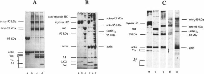
Myosin subfragment-1 (S1) can be cross-linked to two actin monomers by 1-ethyl-3-[3-(dimethylamino)-propyl]-carbodiimide only when F-actin is in excess over S1. Electron micrographs of the covalent actin2-S1 complex showed that S1 was cross-linked to two adjacent monomers of the same actin filament. Cross-linking experiments with pre-proteolyzed S1 derivatives in combination with a proteolytic dissection of the intact covalent actin2-S1 adduct (m = 265 kDa), revealed that two N-terminal segments of actin (residues 1-28) were covalently attached to a single S1 molecule. One was cross-linked to either the 20-kDa or the 50-kDa heavy chain fragments of S1, and the other only to the 50-kDa region. The doubly cross-linked product was formed under physiological ionic strength with S1 or with reconstituted myosin filaments, regardless of the presence of ADP or the regulatory proteins, tropomyosin and troponin. Finally, we found that this cross-linking could also take place within myofibrils in the rigor state. These results demonstrate that under nonsaturating conditions, the actin-S1 interface encompasses a much larger region than that recently proposed for the nonphysiological, fully saturated actin filaments.

摘要

只有当F-肌动蛋白的量超过肌球蛋白亚片段-1(S1)时,1-乙基-3-[3-(二甲基氨基)丙基]-碳二亚胺才能将S1与两个肌动蛋白单体交联。共价肌动蛋白2-S1复合物的电子显微镜照片显示,S1与同一肌动蛋白丝的两个相邻单体交联。用预先蛋白酶解的S1衍生物进行交联实验,并结合对完整共价肌动蛋白2-S1加合物(m = 265 kDa)的蛋白酶解分析,结果表明,肌动蛋白的两个N端片段(第1-28位氨基酸残基)共价连接到单个S1分子上。其中一个片段与S1的20 kDa或50 kDa重链片段交联,另一个仅与50 kDa区域交联。无论是否存在ADP或调节蛋白原肌球蛋白和肌钙蛋白,在生理离子强度下,S1或重组肌球蛋白丝均可形成双交联产物。最后,我们发现这种交联也可以在处于僵直状态的肌原纤维内发生。这些结果表明,在非饱和条件下,肌动蛋白-S1界面所涵盖的区域比最近针对非生理性、完全饱和的肌动蛋白丝所提出的区域要大得多。

https://cdn.ncbi.nlm.nih.gov/pmc/blobs/1a8f/1281859/a7756ddcb43f/biophysj00062-0053-a.jpg
https://cdn.ncbi.nlm.nih.gov/pmc/blobs/1a8f/1281859/62fb98031a35/biophysj00062-0049-a.jpg
https://cdn.ncbi.nlm.nih.gov/pmc/blobs/1a8f/1281859/2b6a637cc508/biophysj00062-0050-a.jpg
https://cdn.ncbi.nlm.nih.gov/pmc/blobs/1a8f/1281859/526af82f3573/biophysj00062-0051-a.jpg
https://cdn.ncbi.nlm.nih.gov/pmc/blobs/1a8f/1281859/a7756ddcb43f/biophysj00062-0053-a.jpg

相似文献

1
A single myosin head can be cross-linked to the N termini of two adjacent actin monomers.单个肌球蛋白头部可以与两个相邻肌动蛋白单体的N端交联。
Biophys J. 1995 Apr;68(4 Suppl):35S-43S.
2
A myosin head can interact with two chemically modified G-actin monomers at ATP-modulated multiple sites.肌球蛋白头部可在ATP调节的多个位点与两个化学修饰的G-肌动蛋白单体相互作用。
Biochemistry. 1996 Dec 17;35(50):16061-8. doi: 10.1021/bi960803s.
3
Mechanism of formation of actomyosin interface.肌动球蛋白界面的形成机制。
J Mol Biol. 2007 Jan 19;365(3):551-4. doi: 10.1016/j.jmb.2006.10.014. Epub 2006 Oct 11.
4
X-ray diffraction evidence for the lack of stereospecific protein interactions in highly activated actomyosin complex.X射线衍射证据表明,在高度活化的肌动球蛋白复合物中不存在立体特异性蛋白质相互作用。
J Mol Biol. 2001 Jan 26;305(4):863-74. doi: 10.1006/jmbi.2000.4334.
5
The role of three-state docking of myosin S1 with actin in force generation.肌球蛋白S1与肌动蛋白的三态对接在力产生中的作用。
Biophys J. 1995 Apr;68(4 Suppl):194S-199S; discussion 199S-201S.
6
Rigor cross-bridges bind to two actin monomers in thin filaments of rabbit psoas muscle.强直横桥与兔腰大肌细肌丝中的两个肌动蛋白单体结合。
J Mol Biol. 1995 Apr 28;248(2):294-307.
7
Probing the hydrophobic interactions in the skeletal actomyosin subfragment 1 and its nucleotide complexes by zero-length cross-linking with a nickel-peptide chelate.通过镍肽螯合物的零长度交联探究骨骼肌肌动球蛋白亚片段1及其核苷酸复合物中的疏水相互作用。
Biochemistry. 1997 Aug 12;36(32):9703-14. doi: 10.1021/bi970615h.
8
Effect of ATP analogues on the actin-myosin interface.ATP类似物对肌动蛋白-肌球蛋白界面的影响。
Biochemistry. 1998 Jun 9;37(23):8385-94. doi: 10.1021/bi980139a.
9
Tropomyosin inhibits the glutaraldehyde-induced cross-link between the central 48-kDa fragment of myosin head and segment 48-67 in actin subdomain 2.原肌球蛋白抑制戊二醛诱导的肌球蛋白头部中央48 kDa片段与肌动蛋白亚结构域2中48 - 67片段之间的交联。
Biochemistry. 1994 Mar 8;33(9):2594-603. doi: 10.1021/bi00175a031.
10
Cooperative rigor binding of myosin to actin is a function of F-actin structure.肌球蛋白与肌动蛋白的协同严格结合是F-肌动蛋白结构的一种功能。
J Mol Biol. 1997 Feb 7;265(5):469-74. doi: 10.1006/jmbi.1996.0761.

引用本文的文献

1
Highly selective inhibition of myosin motors provides the basis of potential therapeutic application.肌球蛋白马达的高度选择性抑制为潜在的治疗应用提供了基础。
Proc Natl Acad Sci U S A. 2016 Nov 22;113(47):E7448-E7455. doi: 10.1073/pnas.1609342113. Epub 2016 Nov 4.
2
Unique charge distribution in surface loops confers high velocity on the fast motor protein Chara myosin.表面环中的独特电荷分布赋予快速运动蛋白 Chara 肌球蛋白高速运动能力。
Proc Natl Acad Sci U S A. 2009 Dec 22;106(51):21585-90. doi: 10.1073/pnas.0910787106. Epub 2009 Dec 2.
3
Binding assay and preliminary X-ray crystallographic analysis of ACTIBIND, a protein with anticarcinogenic and antiangiogenic activities.

本文引用的文献

1
Photochemical cross-linking of the skeletal myosin head heavy chain to actin subdomain-1 at Arg95 and Arg28.
Eur J Biochem. 1993 May 1;213(3):1243-54. doi: 10.1111/j.1432-1033.1993.tb17875.x.
2
Charge-reversion mutagenesis of Dictyostelium actin to map the surface recognized by myosin during ATP-driven sliding motion.对盘基网柄菌肌动蛋白进行电荷反转诱变,以绘制在ATP驱动的滑动运动过程中肌球蛋白识别的表面图谱。
Proc Natl Acad Sci U S A. 1993 Mar 15;90(6):2127-31. doi: 10.1073/pnas.90.6.2127.
3
Interaction and polymerization of the G-actin-myosin head complex: effect of DNase I.G-肌动蛋白-肌球蛋白头部复合体的相互作用与聚合:脱氧核糖核酸酶I的作用
具有抗癌和抗血管生成活性的蛋白质ACTIBIND的结合测定及初步X射线晶体学分析。
Acta Crystallogr Sect F Struct Biol Cryst Commun. 2007 Aug 1;63(Pt 8):716-9. doi: 10.1107/S1744309107034483. Epub 2007 Jul 28.
4
Interaction of myosin with F-actin: time-dependent changes at the interface are not slow.肌球蛋白与F-肌动蛋白的相互作用:界面处随时间的变化并不缓慢。
Biophys J. 2000 Jun;78(6):3093-102. doi: 10.1016/S0006-3495(00)76846-9.
5
Histidine-tagged wild-type yeast actin: its properties and use in an approach for obtaining yeast actin mutants.组氨酸标签野生型酵母肌动蛋白:其特性及在获得酵母肌动蛋白突变体方法中的应用。
Proc Natl Acad Sci U S A. 1999 Mar 16;96(6):2823-7. doi: 10.1073/pnas.96.6.2823.
6
Small segmental rearrangements in the myosin head can explain force generation in muscle.肌球蛋白头部的小片段重排可以解释肌肉中的力产生。
Biophys J. 1996 Aug;71(2):576-89. doi: 10.1016/S0006-3495(96)79292-5.
Biochemistry. 1993 Sep 28;32(38):10005-14. doi: 10.1021/bi00089a016.
4
A structural and kinetic study on myofibrils prevented from shortening by chemical cross-linking.关于通过化学交联阻止肌原纤维缩短的结构和动力学研究。
Biochemistry. 1993 Jul 20;32(28):7255-63. doi: 10.1021/bi00079a023.
5
Three-dimensional atomic model of F-actin decorated with Dictyostelium myosin S1.用盘基网柄菌肌球蛋白S1装饰的F-肌动蛋白的三维原子模型。
Nature. 1993 Jul 8;364(6433):171-4. doi: 10.1038/364171a0.
6
Structure of the actin-myosin complex and its implications for muscle contraction.肌动蛋白-肌球蛋白复合物的结构及其对肌肉收缩的影响。
Science. 1993 Jul 2;261(5117):58-65. doi: 10.1126/science.8316858.
7
Structure of the 265-kilodalton complex formed upon EDC cross-linking of subfragment 1 to F-actin.亚片段1与F-肌动蛋白经EDC交联后形成的265千道尔顿复合物的结构。
Biochemistry. 1993 Dec 21;32(50):13956-60. doi: 10.1021/bi00213a027.
8
Refinement of the F-actin model against X-ray fiber diffraction data by the use of a directed mutation algorithm.通过使用定向突变算法,根据X射线纤维衍射数据对F-肌动蛋白模型进行优化。
J Mol Biol. 1993 Dec 5;234(3):826-36. doi: 10.1006/jmbi.1993.1628.
9
Polarization of fluorescently labeled myosin subfragment-1 fully or partially decorating muscle fibers and myofibrils.荧光标记的肌球蛋白亚片段-1完全或部分修饰肌纤维和肌原纤维的极化。
Biophys J. 1993 Sep;65(3):1027-38. doi: 10.1016/S0006-3495(93)81161-5.
10
Two different rigor complexes of myosin subfragment 1 and actin.
Biochemistry. 1993 Nov 16;32(45):12046-53. doi: 10.1021/bi00096a015.