O'Byrne K J, Ennis J T, Freyne P J, Clancy L J, Prichard J S, Carney D N
Department of Oncology, Mater Misericordiae Hospital, Dublin, Ireland.
Br J Cancer. 1994 Apr;69(4):762-6. doi: 10.1038/bjc.1994.144.
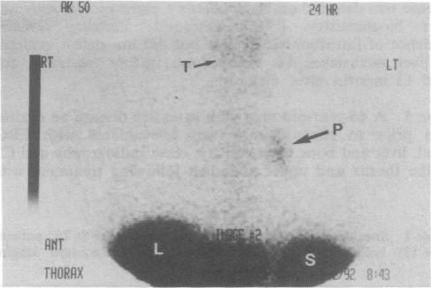
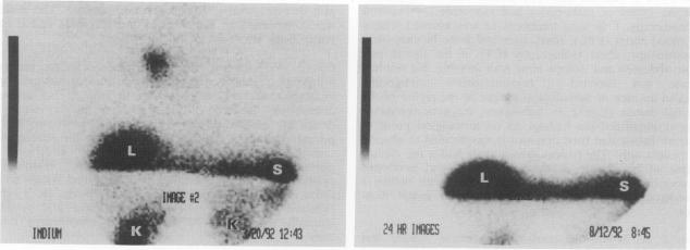
Recent work suggests that between 50 and 75% of small-cell lung cancer (SCLC) tumours have specific high-affinity binding sites for somatostatin. This study evaluated the potential role of the radiolabelled somatostatin analogue, [111In]pentetreotide, in the detection and staging of SCLC in patients prior to and after chemotherapy using scintigraphic imaging techniques. Thirteen patients were studied prior to chemotherapy. Following standard staging six patients had limited stage disease and seven extensive disease. [111In]pentetreotide imaging led to the detection of all primary sites of disease, including a primary site of disease not detectable with chest radiograph or computerised tomography (CT) of the thorax. Five of ten metastatic sites detected by standard staging were also imaged. Furthermore, a cerebellar metastasis was detected in a patient thought to have disease confined to the right hemithorax. This was subsequently confirmed with a CT brain scan. Following chemotherapy [111In]pentetreotide imaging detected residual intrathoracic disease in two of three patients with complete remissions by standard staging and in two patients who had had a partial response to chemotherapy. These results suggest that [111In]pentetreotide imaging may have a role to play in the clinical evaluation of patients with SCLC. Specifically, this technique may be of particular value in detecting residual intrathoracic disease in patients thought to be in complete remission by conventional staging methods.
近期研究表明,50%至75%的小细胞肺癌(SCLC)肿瘤对生长抑素有特定的高亲和力结合位点。本研究采用闪烁成像技术,评估了放射性标记的生长抑素类似物[111In]喷曲肽在化疗前后小细胞肺癌患者检测及分期中的潜在作用。对13例患者在化疗前进行了研究。经过标准分期,6例患者为局限期疾病,7例为广泛期疾病。[111In]喷曲肽成像检测到了所有疾病的原发部位,包括胸部X线片或胸部计算机断层扫描(CT)未检测到的原发部位。标准分期检测到的10个转移部位中有5个也通过成像显示。此外,一名被认为疾病局限于右半胸的患者检测到小脑转移。随后脑部CT扫描证实了这一情况。化疗后,[111In]喷曲肽成像在3例经标准分期完全缓解的患者中的2例以及2例对化疗有部分反应的患者中检测到胸腔内残留疾病。这些结果表明,[111In]喷曲肽成像可能在小细胞肺癌患者的临床评估中发挥作用。具体而言,该技术在检测经传统分期方法认为完全缓解的患者的胸腔内残留疾病方面可能具有特殊价值。