Yao K S, Xanthoudakis S, Curran T, O'Dwyer P J
Fox Chase Cancer Center, Philadelphia, Pennsylvania 19111.
Mol Cell Biol. 1994 Sep;14(9):5997-6003. doi: 10.1128/mcb.14.9.5997-6003.1994.
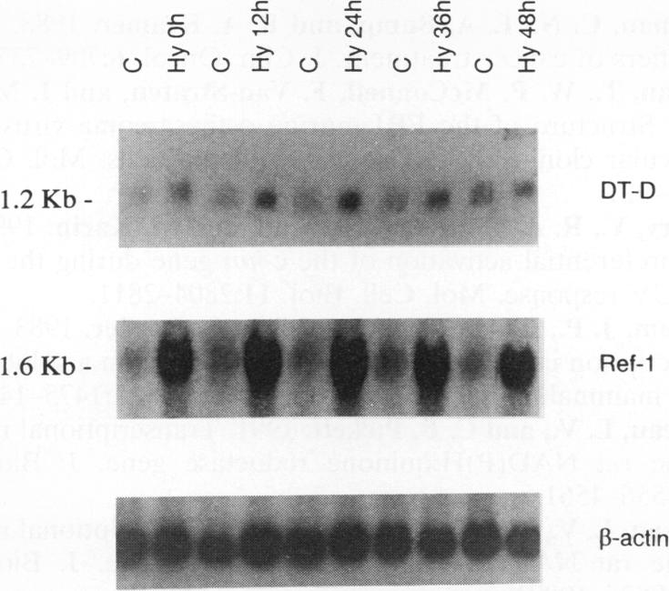
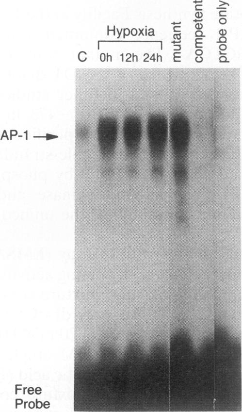
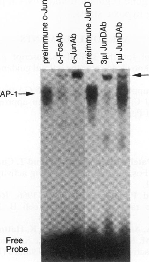
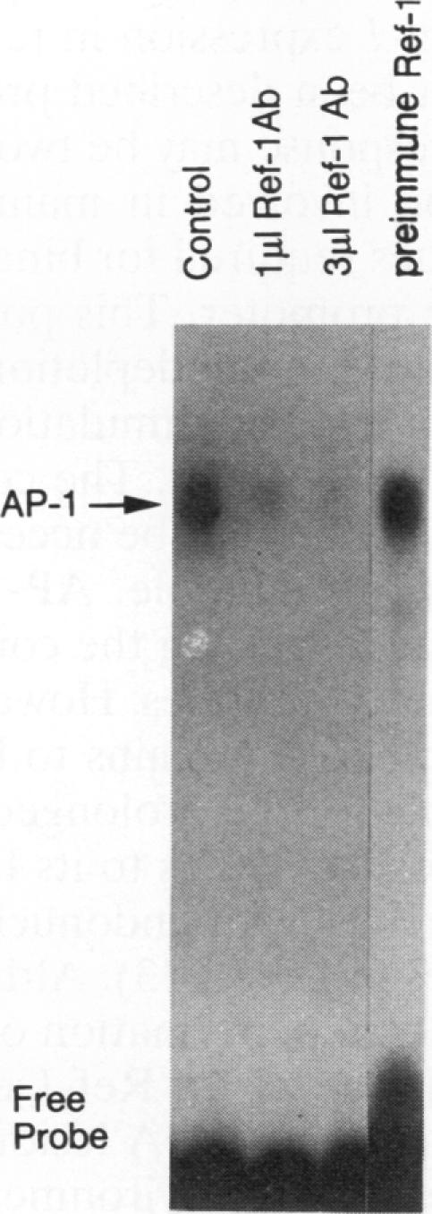
Many solid tumors contain substantial fractions of hypoxic cells which are relatively resistant to both radiation therapy and certain cytotoxic drugs. We have previously shown that exposure of human HT29 cells to hypoxic conditions results in the overexpression of certain enzymes involved in the detoxication of xenobiotics, including NAD(P)H:(quinone acceptor) oxidoreductase (DT)-diaphorase, and gamma-glutamylcysteine synthetase, the rate-limiting enzyme in glutathione synthesis. This hypoxic effect on DT-diaphorase was shown to involve both transcriptional induction and altered message stability. We have investigated the effects of hypoxia on elements in the promoter region of DT-diaphorase. Electrophoretic mobility shift assays demonstrate the induction of a binding activity to the AP-1 response element of DT-diaphorase. Supershift assays suggest that this binding is due to AP-1 nuclear factors and that members of the jun family are induced to a greater degree than fos by hypoxia. Analysis of the kinetics of transcription factor expression indicates that the expression of c-jun and junD is induced during hypoxic exposure; mRNA levels fall during reoxygenation. Induction of fos on the other hand is not as florid during hypoxia (5-fold) and is most pronounced (17-fold) 24 h after the restoration of an oxic environment. Thus, the hypoxic response of DT-diaphorase expression is mediated in part through AP-1, initially by a jun-related mechanism and then by the involvement of fos. The affinity of transcription factors for the AP-1 binding site depends on the redox state of a cysteine residue located close to the DNA-binding region of both Fos and Jun. A nuclear protein, Ref-1, maintains the reduced state of Fos and Jun and promotes binding to AP-1. Nuclear extracts of HT29 cells exposed to hypoxia show markedly increased Ref-1 protein content. Elevation of ref-1 steady-state mRNA levels occurs as an early event following induction of hypoxia and persists when cells are restored to a normally oxygenated environment. Nuclear run-on analysis demonstrates that induction of transcription is the mechanism of ref-1 mRNA elevation. Electrophoretic mobility shift assays and immunodepletion assays were used to further define the interaction of Ref-1 with specific AP-1-binding proteins under hypoxic conditions. These data demonstrate that the induction of detoxicating enzyme expression in HT29 cells exposed to hypoxia results from the induction of both transactivating factors that bind to the AP-1 element and of redox proteins that enhance their affinity for this element.
许多实体瘤含有相当比例的缺氧细胞,这些细胞对放射治疗和某些细胞毒性药物具有相对抗性。我们先前已经表明,将人HT29细胞暴露于缺氧条件下会导致某些参与外源性物质解毒的酶过度表达,包括NAD(P)H:(醌受体)氧化还原酶(DT)-黄递酶,以及γ-谷氨酰半胱氨酸合成酶,后者是谷胱甘肽合成中的限速酶。这种对DT-黄递酶的缺氧效应被证明涉及转录诱导和信息稳定性的改变。我们研究了缺氧对DT-黄递酶启动子区域元件的影响。电泳迁移率变动分析表明,对DT-黄递酶的AP-1反应元件的结合活性被诱导。超迁移分析表明,这种结合是由于AP-1核因子,并且缺氧诱导jun家族成员的程度比fos更大。转录因子表达动力学分析表明,c-jun和junD的表达在缺氧暴露期间被诱导;复氧期间mRNA水平下降。另一方面,fos的诱导在缺氧期间不那么显著(5倍),并且在有氧环境恢复后24小时最为明显(17倍)。因此,DT-黄递酶表达的缺氧反应部分通过AP-1介导,最初通过与jun相关的机制,然后通过fos的参与。转录因子对AP-1结合位点的亲和力取决于位于Fos和Jun的DNA结合区域附近的半胱氨酸残基的氧化还原状态。一种核蛋白Ref-1维持Fos和Jun的还原状态并促进与AP-1的结合。暴露于缺氧的HT29细胞的核提取物显示Ref-1蛋白含量明显增加。缺氧诱导后,ref-1稳态mRNA水平的升高是早期事件,并且当细胞恢复到正常氧合环境时持续存在。核转录分析表明,转录诱导是ref-1 mRNA升高的机制。电泳迁移率变动分析和免疫耗尽分析用于进一步确定缺氧条件下Ref-1与特定AP-1结合蛋白的相互作用。这些数据表明,暴露于缺氧的HT29细胞中解毒酶表达的诱导是由于与AP-1元件结合的反式激活因子和增强其对该元件亲和力的氧化还原蛋白的诱导所致。