• 文献检索
  • 文档翻译
  • 深度研究
  • 学术资讯
  • Suppr Zotero 插件Zotero 插件
  • 邀请有礼
  • 套餐&价格
  • 历史记录
应用&插件
Suppr Zotero 插件Zotero 插件浏览器插件Mac 客户端Windows 客户端微信小程序
定价
高级版会员购买积分包购买API积分包
服务
文献检索文档翻译深度研究API 文档MCP 服务
关于我们
关于 Suppr公司介绍联系我们用户协议隐私条款
关注我们

Suppr 超能文献

核心技术专利:CN118964589B侵权必究
粤ICP备2023148730 号-1Suppr @ 2026

文献检索

告别复杂PubMed语法,用中文像聊天一样搜索,搜遍4000万医学文献。AI智能推荐,让科研检索更轻松。

立即免费搜索

文件翻译

保留排版,准确专业,支持PDF/Word/PPT等文件格式,支持 12+语言互译。

免费翻译文档

深度研究

AI帮你快速写综述,25分钟生成高质量综述,智能提取关键信息,辅助科研写作。

立即免费体验

体内分化的单核吞噬细胞的结构。II. 结核分枝杆菌的作用。

The structure of mononuclear phagocytes differentiating in vivo. II. The effect of Mycobacterium tuberculosis.

作者信息

Adams D O

出版信息

Am J Pathol. 1975 Jul;80(1):101-16.

PMID:808135
原文链接:https://pmc.ncbi.nlm.nih.gov/articles/PMC1912829/
Abstract

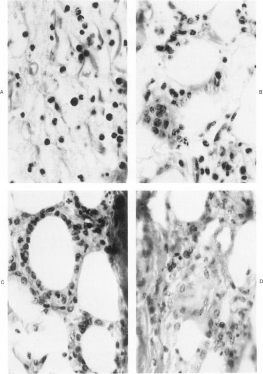
The development and resolution of granulomas induced by Mycobacterium tuberculosis were sequentially traced by correlated light and electron microscopy. The scattered, immature monocytes initially composing the lesions evolved by orderly steps into coalescent, well developed macrophages and ultimately into swirling nests of highly complex epithelioid cells. These ultrastructural changes represent differentiation in vivo of the mononuclear phagocytes. The number of mycobacteria present than waned markedly, and the epithelioid granulomas developed into foreign body granulomas and finally into simple chronic inflammation. Concmonitantly, the epithelioid cells evolved into macrophages and ultimately into immature, monocyte-like forms. These observations suggest that the development of a granuloma represents differntiation in vivo of the constituent mononuclear phagocytes in response to an evoking stimulus. From comparisons with previous studies, mononuclear differentiation in vivo appears to have a fixed pattern and a markedly alterable pace. The observations also suggest a previously undescribed fate for mononuclear phagocytes in developing granulomas. As the granuloma-evoking agent is destroyed, the highly differentiated mononuclear phagocytes change into less mature forms.

摘要

通过相关的光学显微镜和电子显微镜对结核分枝杆菌诱导的肉芽肿的发展和消退进行了连续追踪。最初构成病变的散在、未成熟单核细胞按有序步骤演变为融合的、发育良好的巨噬细胞,最终演变为高度复杂的上皮样细胞的漩涡状巢。这些超微结构变化代表了单核吞噬细胞在体内的分化。随后存在的分枝杆菌数量明显减少,上皮样肉芽肿发展为异物肉芽肿,最终发展为单纯性慢性炎症。与此同时,上皮样细胞演变为巨噬细胞,最终演变为未成熟的、单核细胞样形态。这些观察结果表明,肉芽肿的发展代表了组成单核吞噬细胞在体内对诱发刺激的分化。通过与先前研究的比较,体内单核细胞分化似乎有固定模式和明显可变的速度。这些观察结果还提示了发育中的肉芽肿中单核吞噬细胞以前未描述的命运。随着肉芽肿诱发剂被破坏,高度分化的单核吞噬细胞转变为不太成熟的形式。

https://cdn.ncbi.nlm.nih.gov/pmc/blobs/ba04/1912829/44a520acc2be/amjpathol00458-0121-a.jpg
https://cdn.ncbi.nlm.nih.gov/pmc/blobs/ba04/1912829/0f1e264d8866/amjpathol00458-0122-a.jpg
https://cdn.ncbi.nlm.nih.gov/pmc/blobs/ba04/1912829/0465e018ba9c/amjpathol00458-0120-a.jpg
https://cdn.ncbi.nlm.nih.gov/pmc/blobs/ba04/1912829/44a520acc2be/amjpathol00458-0121-a.jpg
https://cdn.ncbi.nlm.nih.gov/pmc/blobs/ba04/1912829/0f1e264d8866/amjpathol00458-0122-a.jpg
https://cdn.ncbi.nlm.nih.gov/pmc/blobs/ba04/1912829/0465e018ba9c/amjpathol00458-0120-a.jpg
https://cdn.ncbi.nlm.nih.gov/pmc/blobs/ba04/1912829/44a520acc2be/amjpathol00458-0121-a.jpg

相似文献

1
The structure of mononuclear phagocytes differentiating in vivo. II. The effect of Mycobacterium tuberculosis.体内分化的单核吞噬细胞的结构。II. 结核分枝杆菌的作用。
Am J Pathol. 1975 Jul;80(1):101-16.
2
The structure of mononuclear phagocytes differentiating in vivo. I. Sequential fine and histologic studies of the effect of Bacillus Calmette-Guerin (BCG).体内分化的单核吞噬细胞的结构。I. 卡介苗(BCG)作用的连续精细和组织学研究。
Am J Pathol. 1974 Jul;76(1):17-48.
3
The structure of mononuclear phagocytes differentiating in vivo. III. The effect of particulate foreign substances.体内分化的单核吞噬细胞的结构。III. 颗粒状异物的作用。
Am J Pathol. 1977 Nov;89(2):335-50.
4
The origin, morphology, and function of epithelioid cells.上皮样细胞的起源、形态及功能。
Immunobiology. 1982 Apr;161(3-4):274-82. doi: 10.1016/S0171-2985(82)80083-1.
5
Experimental epithelioid cell granulomas, tubercle formation and immunological competence: an ultrastructural analysis.实验性上皮样细胞肉芽肿、结核结节形成与免疫能力:超微结构分析
J Pathol. 1983 Oct;141(2):97-112. doi: 10.1002/path.1711410202.
6
The differentiation of monocytes into macrophages, epithelioid cells, and multinucleated giant cells in subcutaneous granulomas. I. Fine structure.皮下肉芽肿中单核细胞向巨噬细胞、上皮样细胞和多核巨细胞的分化。I. 精细结构
Cell Tissue Res. 1979 Apr 12;197(3):355-78. doi: 10.1007/BF00233563.
7
Experimental granulomatous inflammation: the ultrastructure of the granuloma induced by injection of tubercle bacilli into Freund adjuvant-sensitised guinea-pigs.实验性肉芽肿性炎症:向弗氏佐剂致敏豚鼠注射结核杆菌所诱导肉芽肿的超微结构
J Pathol. 1979 Dec;129(4):191-201. doi: 10.1002/path.1711290405.
8
Experimental mycobacterial granulomas in guinea pig lymph nodes: ultrastructural observations.豚鼠淋巴结中实验性分枝杆菌肉芽肿:超微结构观察
J Pathol. 1981 Aug;134(4):253-65. doi: 10.1002/path.1711340402.
9
In situ ultrastructural detection and quantitation of liver mononuclear phagocytes in contact with hepatocytes in chronic type B hepatitis.慢性乙型肝炎中与肝细胞接触的肝单核吞噬细胞的原位超微结构检测与定量分析
Lab Invest. 1984 Dec;51(6):667-74.
10
Dynamics of mononuclear phagocytes in lymph nodes and granulomas in chronic tuberculous inflammation.慢性结核性炎症中淋巴结和肉芽肿内单核吞噬细胞的动态变化
Bull Exp Biol Med. 2001 Feb;131(2):165-8. doi: 10.1023/a:1017552214329.

引用本文的文献

1
Host Cell Death and Modulation of Immune Response against Infection.宿主细胞死亡与 感染免疫应答的调节。
Int J Mol Sci. 2024 Jun 6;25(11):6255. doi: 10.3390/ijms25116255.
2
Hypoxia Promotes a Mixed Inflammatory-Fibrotic Macrophages Phenotype in Active Sarcoidosis.缺氧促进活动期结节病中混合炎症-纤维化的巨噬细胞表型。
Front Immunol. 2021 Aug 11;12:719009. doi: 10.3389/fimmu.2021.719009. eCollection 2021.
3
CD4+ cell-dependent granuloma formation in humanized mice infected with mycobacteria.在感染分枝杆菌的人源化小鼠中,CD4+ 细胞依赖性肉芽肿形成。

本文引用的文献

1
THE IN VITRO DIFFERENTIATION OF MONONUCLEAR PHAGOCYTES. II. THE INFLUENCE OF SERUM ON GRANULE FORMATION, HYDROLASE PRODUCTION, AND PINOCYTOSIS.单核吞噬细胞的体外分化。II. 血清对颗粒形成、水解酶产生及胞饮作用的影响。
J Exp Med. 1965 May 1;121(5):835-48. doi: 10.1084/jem.121.5.835.
2
[Study by electron microscopy of experimental renal tuberculosis in a resistant animal (rat)].[对耐药动物(大鼠)实验性肾结核的电子显微镜研究]
Rev Tuberc Pneumol (Paris). 1960 Dec;24:1271-84.
3
The cellular evolution of inflammatory granulomata.炎性肉芽肿的细胞演变
Proc Natl Acad Sci U S A. 2013 Apr 16;110(16):6482-7. doi: 10.1073/pnas.1219985110. Epub 2013 Apr 4.
4
Regulation of monocyte differentiation by specific signaling modules and associated transcription factor networks.特定信号模块和相关转录因子网络对单核细胞分化的调控。
Cell Mol Life Sci. 2014 Jan;71(1):63-92. doi: 10.1007/s00018-013-1322-4. Epub 2013 Mar 24.
5
The role of the granuloma in expansion and dissemination of early tuberculous infection.肉芽肿在早期结核感染的扩展与播散中的作用。
Cell. 2009 Jan 9;136(1):37-49. doi: 10.1016/j.cell.2008.11.014.
6
Dynamic nature of host-pathogen interactions in Mycobacterium marinum granulomas.海鱼分枝杆菌肉芽肿中宿主-病原体相互作用的动态特性。
Infect Immun. 2001 Dec;69(12):7820-31. doi: 10.1128/IAI.69.12.7820-7831.2001.
7
Mycobacterium tuberculosis alters expression of adhesion molecules on monocytic cells.结核分枝杆菌会改变单核细胞上黏附分子的表达。
Infect Immun. 1994 Jun;62(6):2515-20. doi: 10.1128/iai.62.6.2515-2520.1994.
8
[Multinucleated giant cells in granulomas. Reorganization of the internal structure after confluence of macrophage system cells (author's transl)].[肉芽肿中的多核巨细胞。巨噬细胞系统细胞融合后内部结构的重组(作者译)]
Virchows Arch A Pathol Anat Histol. 1980;385(3):309-33. doi: 10.1007/BF00432540.
9
Epithelioid granuloma formation by a synthetic bacterial cell wall component, muramyl dipeptide (MDP).由合成细菌细胞壁成分胞壁酰二肽(MDP)形成上皮样肉芽肿。
Am J Pathol. 1980 Mar;98(3):733-48.
10
Experimental pulmonary candidiasis in modified rabbits. Histopathological, ultrastructural and enzyme cytochemical studies of tissue reactions.改良兔实验性肺念珠菌病。组织反应的组织病理学、超微结构和酶细胞化学研究。
Mycopathologia. 1984 Apr 15;85(3):129-44. doi: 10.1007/BF00440942.
J Pathol Bacteriol. 1966 Jul;92(1):163-7. doi: 10.1002/path.1700920117.
4
The in vitro differentiation of mononuclear phagocytes. IV. The ultrastructure of macrophage differentiation in the peritoneal cavity and in culture.单核吞噬细胞的体外分化。IV. 腹腔及培养中巨噬细胞分化的超微结构
J Exp Med. 1966 Apr 1;123(4):747-56. doi: 10.1084/jem.123.4.747.
5
Transformation of monocytes in tissue culture into macrophages, epithelioid cells, and multinucleated giant cells. An electron microscope study.组织培养中单核细胞向巨噬细胞、上皮样细胞和多核巨细胞的转化。一项电子显微镜研究。
J Cell Biol. 1966 Feb;28(2):303-32. doi: 10.1083/jcb.28.2.303.
6
Changes in the fine structure of macrophages in experimentally produced tuberculous granulomas in hamsters.仓鼠实验性结核性肉芽肿中巨噬细胞精细结构的变化
Lab Invest. 1965 Nov;14(11):2034-55.
7
Modification of macrophage function.巨噬细胞功能的改变。
J Reticuloendothel Soc. 1968 Jun;5(3):179-202.
8
The fine structure of sarcoid and tuberculous granulomas.结节病和结核性肉芽肿的精细结构。
Postgrad Med J. 1970 Aug;46(538):496-500. doi: 10.1136/pgmj.46.538.496.
9
Composition, morphology, and source of cells in delayed skin reactions.迟发性皮肤反应中细胞的组成、形态及来源
Am J Pathol. 1970 Feb;58(2):201-18.
10
Macrophage accumulation, division, maturation, and digestive and microbicidal capacities in tuberculous lesions. 3. The turnover of macrophages and its relation to their activation and antimicrobial immunity in primary BCG lesions and those of reinfection.结核病变中巨噬细胞的聚集、分裂、成熟以及消化和杀菌能力。3. 巨噬细胞在原发性卡介苗接种病变和再感染病变中的更新及其与激活和抗微生物免疫的关系。
J Immunol. 1972 Nov;109(5):1109-21.