INSERM UMR 1272, Sorbonne Paris-Nord University, Bobigny, France.
AP-HP, Pulmonology Department, Avicenne Hospital, Bobigny, France.
Front Immunol. 2021 Aug 11;12:719009. doi: 10.3389/fimmu.2021.719009. eCollection 2021.
Macrophages are pivotal cells in sarcoidosis. Monocytes-derived (MD) macrophages have recently been demonstrated to play a major role especially in pulmonary sarcoidosis. From inflammatory tissues to granulomas, they may be exposed to low oxygen tension environments. As hypoxia impact on sarcoidosis immune cells has never been addressed, we designed the present study to investigate MD-macrophages from sarcoidosis patients in this context. We hypothesized that hypoxia may induce functional changes on MD-macrophages which could have a potential impact on the course of sarcoidosis.
We studied MD-macrophages, from high active sarcoidosis (AS) (n=26), low active or inactive sarcoidosis (IS) (n=24) and healthy controls (n=34) exposed 24 hours to normoxia (21% O) or hypoxia (1.5% O). Different macrophage functions were explored: hypoxia-inducible factor-1α (HIF-1α) and nuclear factor-kappa B (NF-κB) activation, cytokines secretion, phagocytosis, CD80/CD86/HLA-DR expression, profibrotic response.
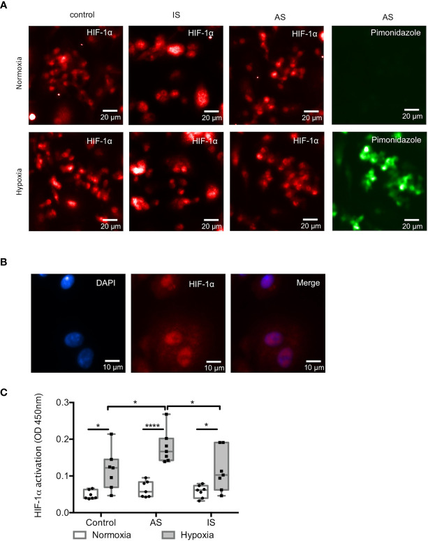
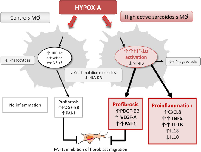
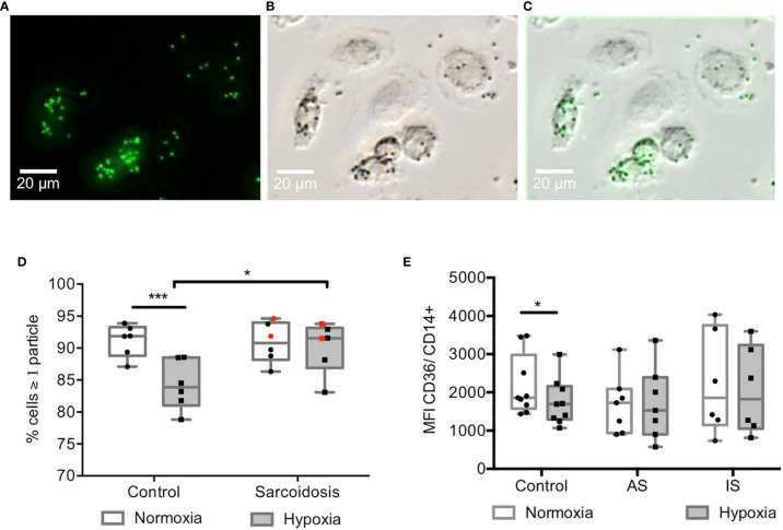
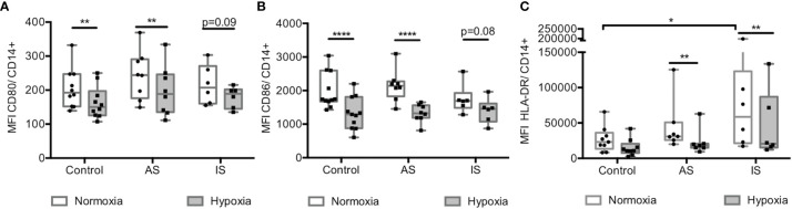
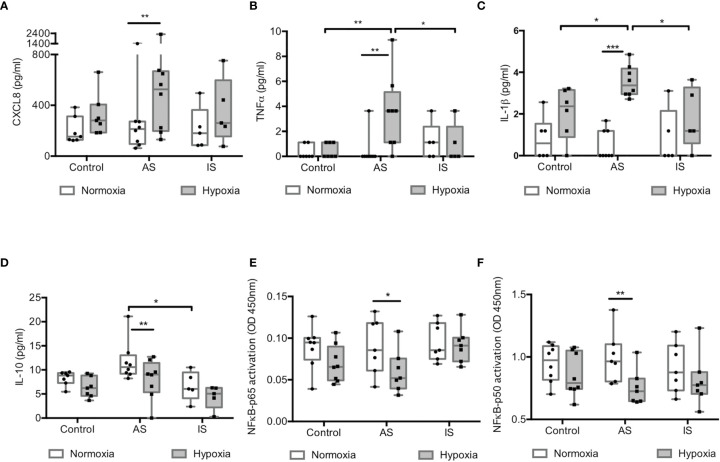
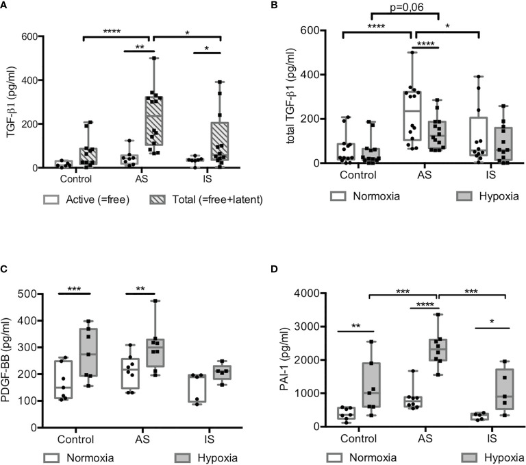
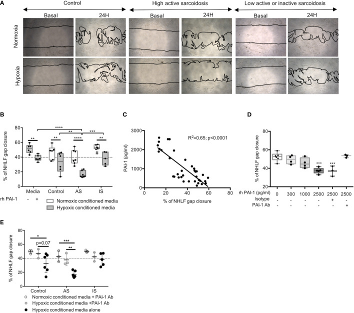
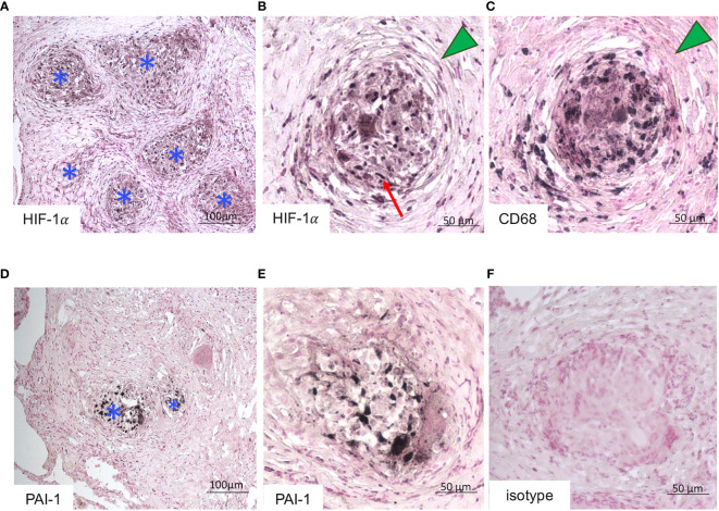
We observed that hypoxia, with a significantly more pronounced effect in AS compared with controls and IS, increased the HIF-1α trans-activity, promoted a proinflammatory response (TNFα, IL1ß) without activating NF-κB pathway and a profibrotic response (TGFß1, PDGF-BB) with PAI-1 secretion associated with human lung fibroblast migration inhibition. These results were confirmed by immunodetection of HIF-1α and PAI-1 in granulomas observed in pulmonary biopsies from patients with sarcoidosis. Hypoxia also decreased the expression of CD80/CD86 and HLA-DR on MD-macrophages in the three groups while it did not impair phagocytosis and the expression of CD36 expression on cells in AS and IS at variance with controls.
Hypoxia had a significant impact on MD-macrophages from sarcoidosis patients, with the strongest effect seen in patients with high active disease. Therefore, hypoxia could play a significant role in sarcoidosis pathogenesis by increasing the macrophage proinflammatory response, maintaining phagocytosis and reducing antigen presentation, leading to a deficient T cell response. In addition, hypoxia could favor fibrosis by promoting profibrotic cytokines response and by sequestering fibroblasts in the vicinity of granulomas.
巨噬细胞是肉样瘤病中的关键细胞。最近已经证明,单核细胞衍生的(MD)巨噬细胞在肺肉样瘤病中发挥着主要作用。从炎症组织到肉芽肿,它们可能会暴露在低氧张力环境中。由于缺氧对肉样瘤病免疫细胞的影响从未得到解决,我们设计了本研究以在这种情况下研究肉样瘤病患者的 MD 巨噬细胞。我们假设缺氧可能会诱导 MD 巨噬细胞发生功能变化,这可能对肉样瘤病的病程产生潜在影响。
我们研究了 26 例高活性肉样瘤病(AS)、24 例低活性或不活跃肉样瘤病(IS)和 34 例健康对照者的 MD 巨噬细胞,这些细胞在 24 小时内暴露于常氧(21% O)或缺氧(1.5% O)环境中。研究了不同的巨噬细胞功能:缺氧诱导因子-1α(HIF-1α)和核因子-κB(NF-κB)的激活、细胞因子的分泌、吞噬作用、CD80/CD86/HLA-DR 的表达、成纤维细胞增殖反应。
我们发现,与对照组和 IS 相比,缺氧在 AS 中具有更显著的作用,增加了 HIF-1α的转录活性,促进了促炎反应(TNFα、IL1β),而没有激活 NF-κB 途径,并导致成纤维细胞增殖反应(TGFβ1、PDGF-BB),同时伴有组织纤溶酶原激活物抑制剂 1(PAI-1)的分泌,这与人类肺成纤维细胞迁移抑制有关。这些结果通过在肺活检中观察到的肉样瘤病患者的肉芽肿中 HIF-1α和 PAI-1 的免疫检测得到了证实。缺氧还降低了三组 MD 巨噬细胞中 CD80/CD86 和 HLA-DR 的表达,但在 AS 和 IS 中不损害吞噬作用和 CD36 的表达,与对照组不同。
缺氧对肉样瘤病患者的 MD 巨噬细胞有显著影响,在高活性疾病患者中影响最大。因此,缺氧可能通过增加巨噬细胞的促炎反应、维持吞噬作用和减少抗原呈递,导致 T 细胞反应不足,从而在肉样瘤病发病机制中发挥重要作用。此外,缺氧通过促进成纤维细胞增殖反应和将成纤维细胞隔离在肉芽肿附近,可能有利于纤维化。