Ovcinnikov N M, Delektorskij V V, Turanova E N, Yashkova G N
Br J Vener Dis. 1975 Dec;51(6):357-75. doi: 10.1136/sti.51.6.357.
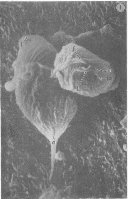
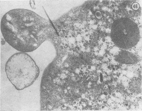
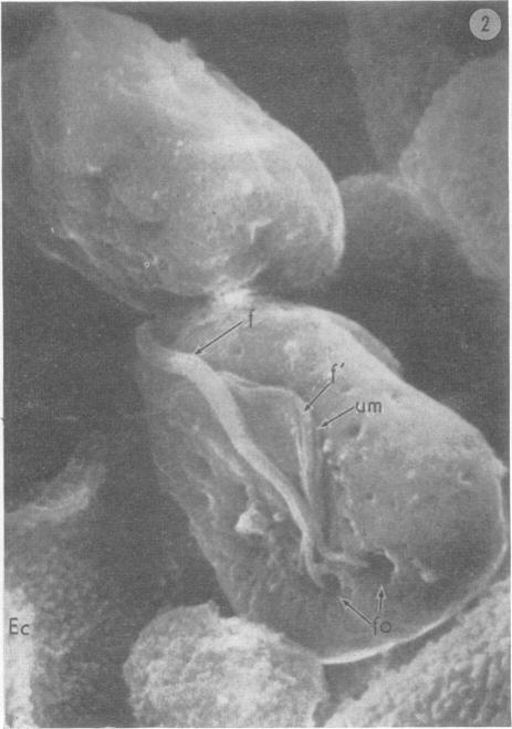
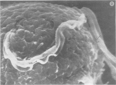
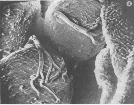
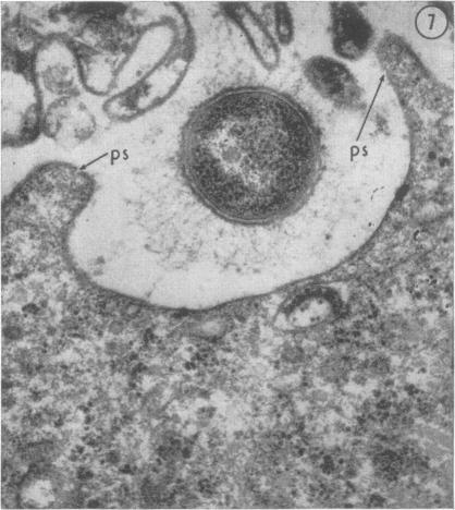
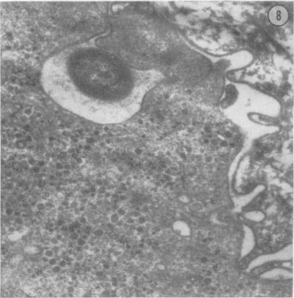
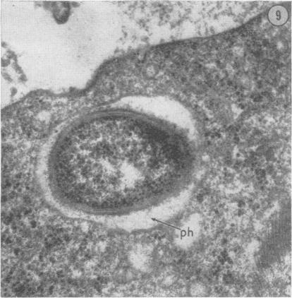
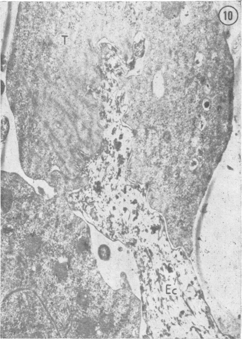
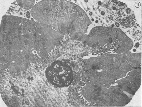
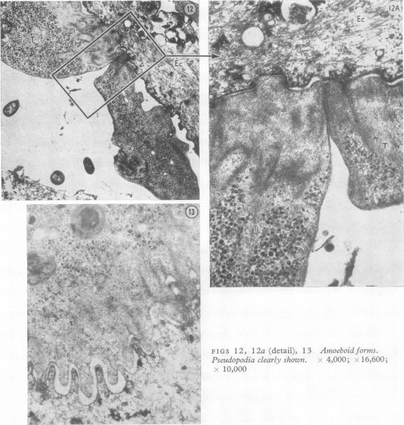
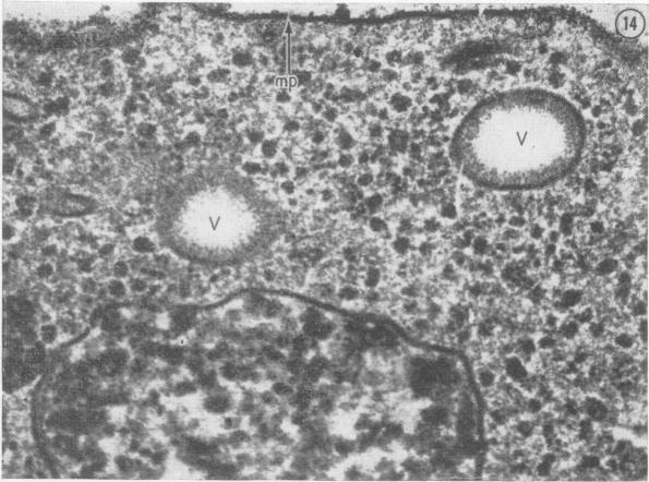
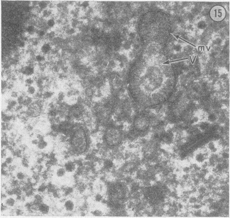
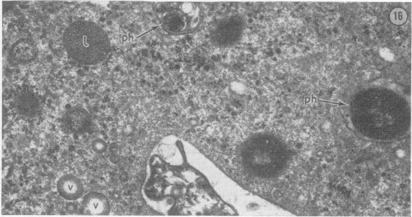
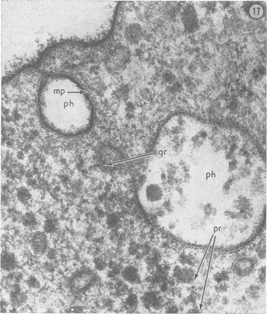
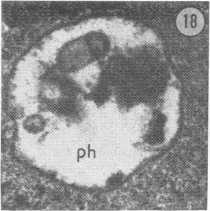
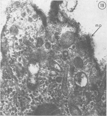
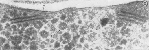
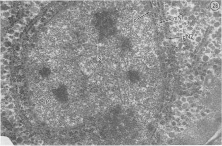
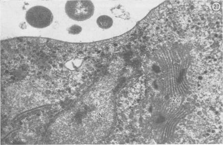
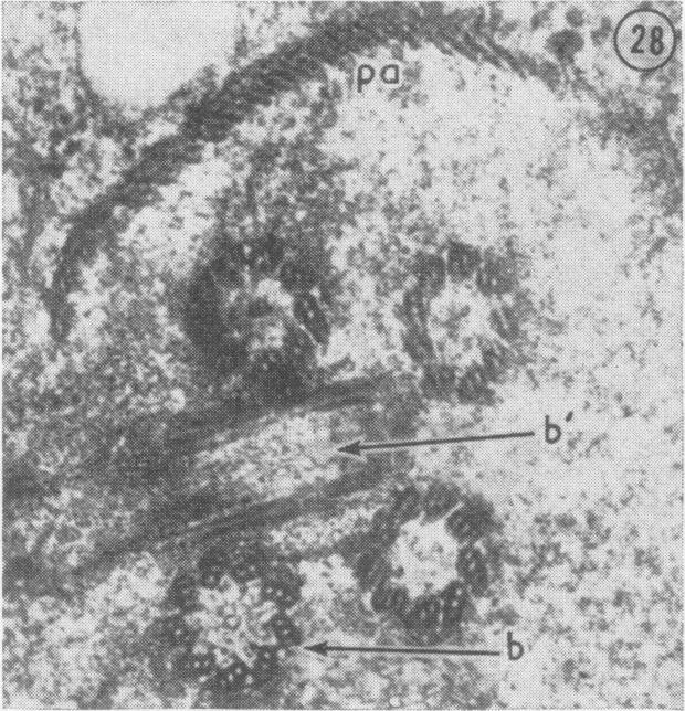
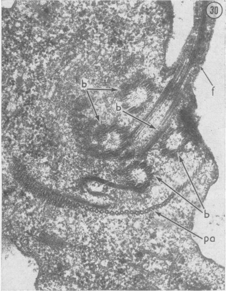
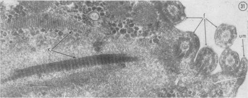
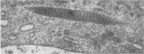
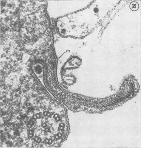
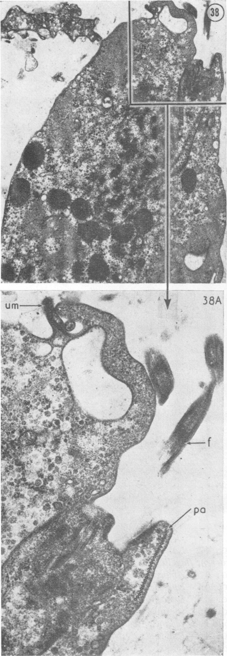
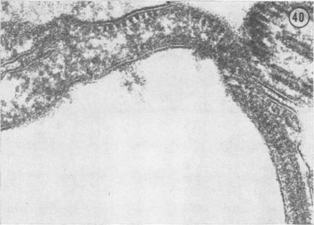
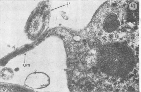
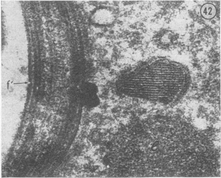
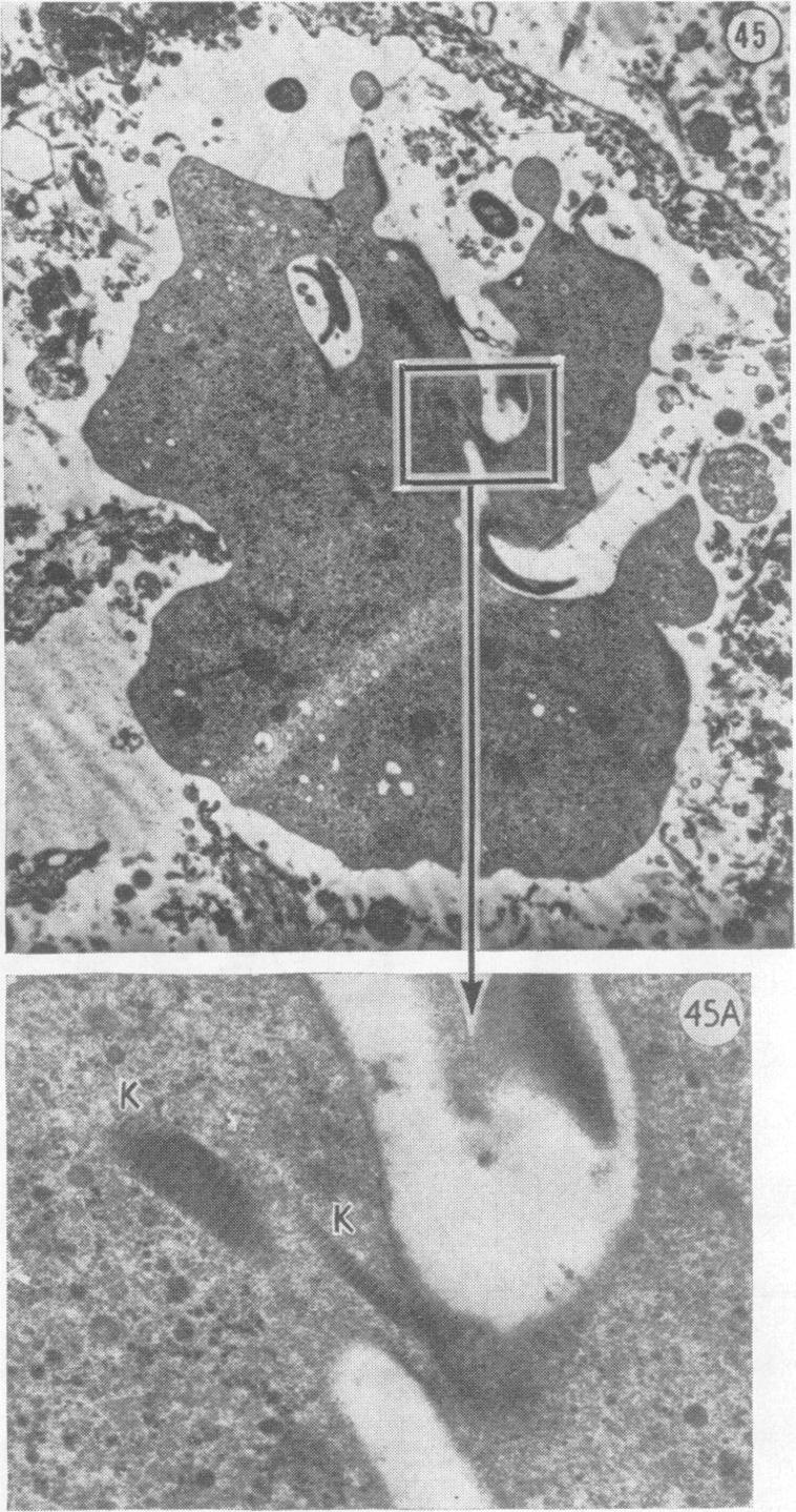
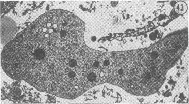
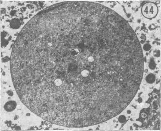
Under the scanning electron microscope, the body surface of trichomonads appears ruffled and creased, with numerous crater-like depressions, which should probably be interpreted as the initial stage in the formation of digestive vacuoles or pinocytotic vesicles. Having contacted epithelial cells, trichomonads engulf them totally or partially. Various micro-organisms also become a prey of the protozoon. The different stages of phagocytosis are illustrated in electron micrographs. Gonococci have been discovered in phagosomes of T. vaginalis. Usually their phagocytosis is not brought to completion and they survive within the trichomonads (endocytobiosis). This suggests that the agent of gonorrhoea may be maintained within trichomonads in cases of mixed infection. In addition to morphological details described earlier, T. vaginalis has lattice-like and lamellar structures of uncertain function. Spherical forms are found usually in an unfavourable environment or as a result of budding.
在扫描电子显微镜下,滴虫的体表呈现出褶皱和皱纹,有许多火山口状的凹陷,这可能应被解释为消化液泡或胞饮小泡形成的初始阶段。与上皮细胞接触后,滴虫会完全或部分吞噬它们。各种微生物也会成为这种原生动物的猎物。吞噬作用的不同阶段在电子显微照片中得到了展示。在阴道毛滴虫的吞噬体中发现了淋球菌。通常它们的吞噬作用不会完成,而是在滴虫内存活(内共生)。这表明在混合感染的情况下,淋病病原体可能在滴虫内得以维持。除了前面描述的形态学细节外,阴道毛滴虫还有功能不明的格子状和层状结构。球形形态通常在不利环境中出现或通过出芽形成。