Tsuchida M, Hanawa H, Hirahara H, Watanabe H, Matsumoto Y, Sekikawa H, Abo T
Department of Immunology, Niigata University School of Medicine, Japan.
Immunology. 1994 Mar;81(3):420-7.
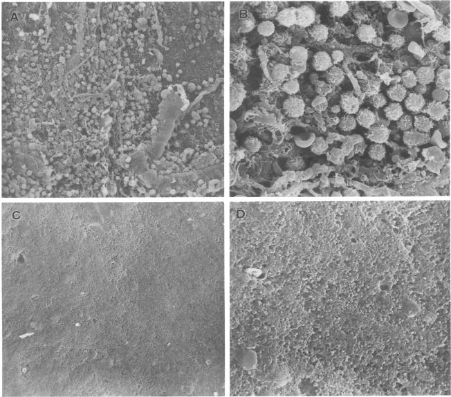
Experimental autoimmune encephalomyelitis (EAE) was induced in Lewis rats to elucidate the origin of effector T cells and the route by which they invade lesions. Since mouse studies have suggested that some autoimmune diseases are induced by extrathymic T cells in the liver, we focused our attention on the properties of mononuclear cells (MNC) isolated from the liver and other organs in rats with EAE. A small but significant proportion of LFA-1+ alpha beta T cells was identified in the liver as early as day 7 after immunization with myelin basic protein (MBP). Such LFA-1+ alpha beta T cells were also abundant among MNC attached to the spinal cord (i.e. subarachnoid space), and MNC infiltrated the spinal cord in rats with EAE (day 12). In electron microscopy, MNC attached to the spinal cord were found to be quite unique in terms of their large cell size with well-developed microvilli. More importantly, they were comprised of a considerably large proportion of double-negative CD4- CD8- T cells as well as single-positive CD4+ T cells. However, the cells which infiltrated the spinal cord were mainly CD4+. The present results raise the possibility that the subarachnoid space might be a major site for the expansion of extrathymic T cells in rats with EAE, and that only a limited population of CD4+ T cells invade the spinal cord directly through the outer layer and elicit EAE.
为了阐明效应T细胞的起源及其侵入病变的途径,我们在Lewis大鼠中诱导了实验性自身免疫性脑脊髓炎(EAE)。由于小鼠研究表明,一些自身免疫性疾病是由肝脏中的胸腺外T细胞诱导的,因此我们将注意力集中在从患有EAE的大鼠的肝脏和其他器官中分离出的单核细胞(MNC)的特性上。早在用髓鞘碱性蛋白(MBP)免疫后第7天,就在肝脏中鉴定出一小部分但比例显著的LFA-1+αβT细胞。在附着于脊髓(即蛛网膜下腔)的MNC中,这种LFA-1+αβT细胞也很丰富,并且在患有EAE的大鼠(第12天)中,MNC浸润脊髓。在电子显微镜下,发现附着于脊髓的MNC在细胞体积大且微绒毛发达方面非常独特。更重要的是,它们由相当大比例的双阴性CD4-CD8-T细胞以及单阳性CD4+T细胞组成。然而,浸润脊髓的细胞主要是CD4+。目前的结果提出了一种可能性,即蛛网膜下腔可能是患有EAE的大鼠中胸腺外T细胞扩增的主要部位,并且只有有限数量的CD4+T细胞直接通过外层侵入脊髓并引发EAE。