• 文献检索
  • 文档翻译
  • 深度研究
  • 学术资讯
  • Suppr Zotero 插件Zotero 插件
  • 邀请有礼
  • 套餐&价格
  • 历史记录
应用&插件
Suppr Zotero 插件Zotero 插件浏览器插件Mac 客户端Windows 客户端微信小程序
定价
高级版会员购买积分包购买API积分包
服务
文献检索文档翻译深度研究API 文档MCP 服务
关于我们
关于 Suppr公司介绍联系我们用户协议隐私条款
关注我们

Suppr 超能文献

核心技术专利:CN118964589B侵权必究
粤ICP备2023148730 号-1Suppr @ 2026

文献检索

告别复杂PubMed语法,用中文像聊天一样搜索,搜遍4000万医学文献。AI智能推荐,让科研检索更轻松。

立即免费搜索

文件翻译

保留排版,准确专业,支持PDF/Word/PPT等文件格式,支持 12+语言互译。

免费翻译文档

深度研究

AI帮你快速写综述,25分钟生成高质量综述,智能提取关键信息,辅助科研写作。

立即免费体验

伤口角质形成细胞中整合素和基底膜成分的表达。

Expression of integrins and basement membrane components by wound keratinocytes.

作者信息

Larjava H, Salo T, Haapasalmi K, Kramer R H, Heino J

机构信息

Department of Periodontology, University of Turku, Finland.

出版信息

J Clin Invest. 1993 Sep;92(3):1425-35. doi: 10.1172/JCI116719.

DOI:10.1172/JCI116719
PMID:8376596
原文链接:https://pmc.ncbi.nlm.nih.gov/articles/PMC288287/
Abstract

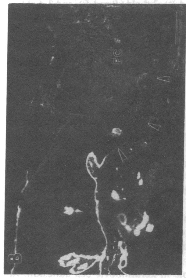
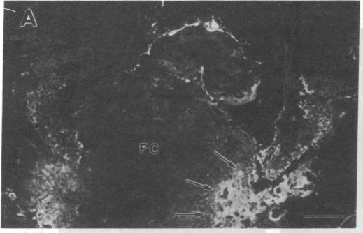
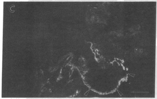
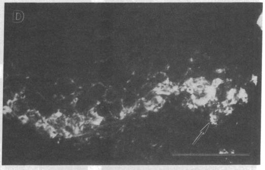
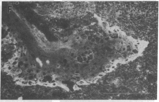
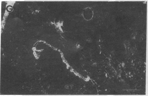
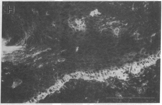
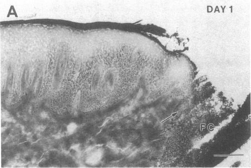
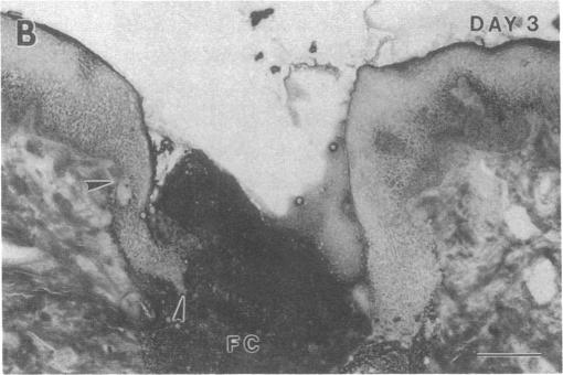
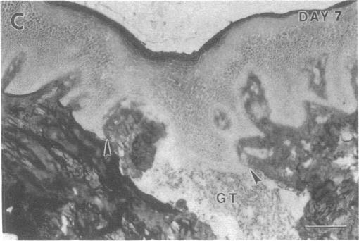
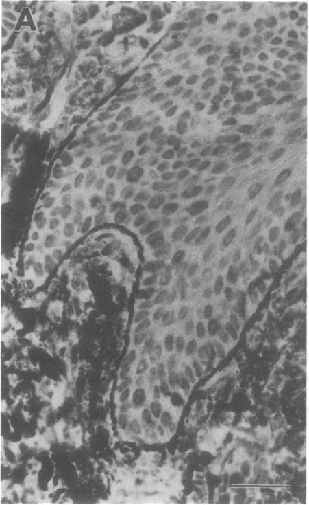
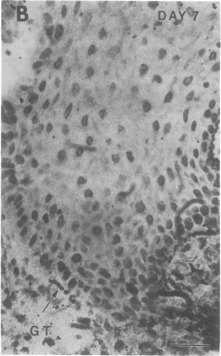
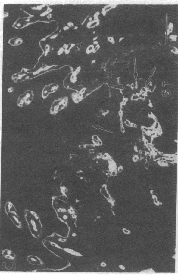
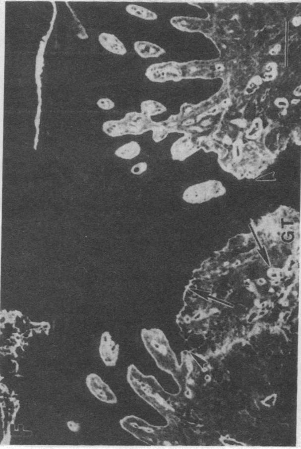
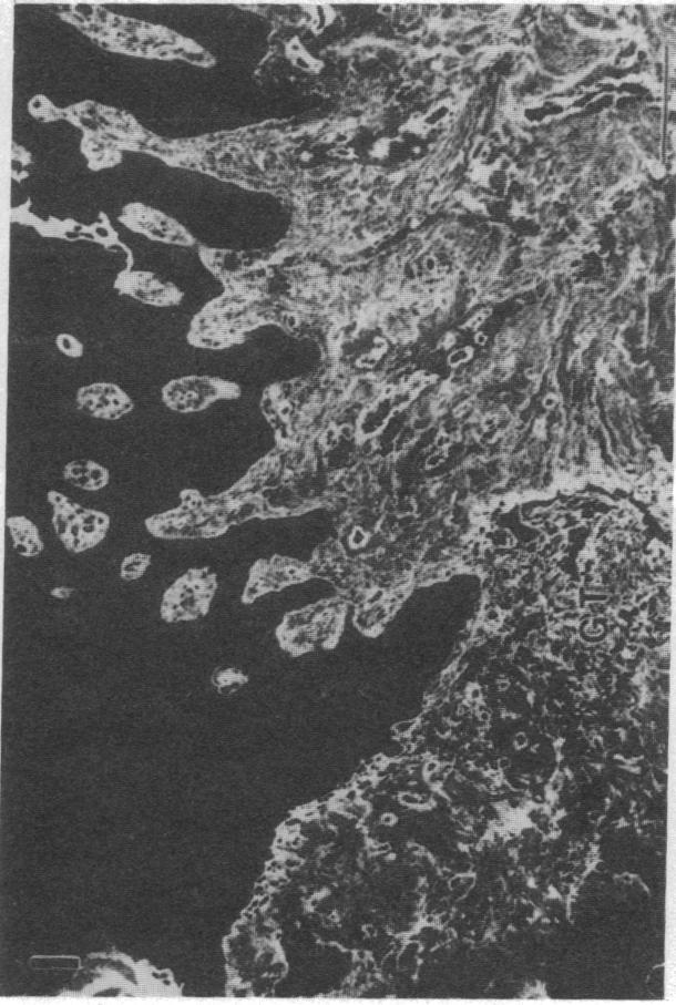
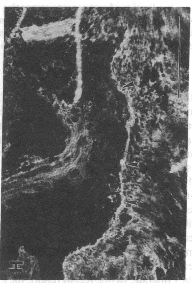
Extracellular matrix proteins and their cellular receptors, integrins, play a fundamental role in keratinocyte adhesion and migration. During wound healing, keratinocytes detach, migrate until the two epithelial sheets confront, and then regenerate the basement membrane. We examined the expression of different integrins and their putative ligands in keratinocytes during human mucosal wound healing. Migrating keratinocytes continuously expressed kalinin but not the other typical components of the basement membrane zone: type IV collagen, laminin, and type VII collagen. When the epithelial sheets confronted each other, these missing basement membrane components started to appear gradually through the entire wound area. The expression of integrin beta 1 subunit was increased in keratinocytes during migration. The beta 1-associated alpha 2 and alpha 3 subunits were expressed constantly by wound keratinocytes whereas the alpha 5 subunit was present only in keratinocytes during reepithelialization. Furthermore, migrating cells started to express alpha v-integrins which were not present in the nonaffected epithelium. All keratinocytes also expressed the alpha 6 beta 4 integrin during migration. In the migrating cells, the distribution of integrins was altered. In normal mucosa, beta 1-integrins were located mainly on the lateral plasma membrane and alpha 6 beta 4 at the basal surface of basal keratinocytes in the nonaffected tissue. In wounds, integrins were found in filopodia of migrating keratinocytes, and also surrounding cells in several cell layers of the migrating sheet. The results indicate that migrating keratinocytes, in deep human wounds enlarge their integrin repertoire. The changes in integrin expression take place concomitantly with changes in the basement membrane composition, suggesting a close interplay of these two groups of molecules during wound healing.

摘要

细胞外基质蛋白及其细胞受体整合素在角质形成细胞的黏附和迁移中起重要作用。在伤口愈合过程中,角质形成细胞脱离、迁移,直到两个上皮层相遇,然后再生基底膜。我们研究了人类黏膜伤口愈合过程中角质形成细胞中不同整合素及其假定配体的表达情况。迁移的角质形成细胞持续表达角蛋白,但不表达基底膜区的其他典型成分:IV型胶原蛋白、层粘连蛋白和VII型胶原蛋白。当上皮层相互面对时,这些缺失的基底膜成分开始逐渐在整个伤口区域出现。整合素β1亚基在角质形成细胞迁移过程中的表达增加。与β1相关的α2和α3亚基由伤口角质形成细胞持续表达,而α5亚基仅在上皮再形成过程中的角质形成细胞中存在。此外,迁移细胞开始表达在未受影响的上皮中不存在的αv整合素。所有角质形成细胞在迁移过程中也表达α6β4整合素。在迁移细胞中,整合素的分布发生了改变。在正常黏膜中,β1整合素主要位于侧面质膜上,α6β4位于未受影响组织中基底角质形成细胞的基底表面。在伤口中,整合素存在于迁移角质形成细胞的丝状伪足中,也存在于迁移层的几个细胞层中的周围细胞中。结果表明,在人类深部伤口中迁移的角质形成细胞扩大了其整合素种类。整合素表达的变化与基底膜组成的变化同时发生,这表明在伤口愈合过程中这两组分子之间存在密切的相互作用。

https://cdn.ncbi.nlm.nih.gov/pmc/blobs/c843/288287/5a62e305bcf0/jcinvest00041-0325-f.jpg
https://cdn.ncbi.nlm.nih.gov/pmc/blobs/c843/288287/126d1fc8408d/jcinvest00041-0328-a.jpg
https://cdn.ncbi.nlm.nih.gov/pmc/blobs/c843/288287/04a2062614d1/jcinvest00041-0328-b.jpg
https://cdn.ncbi.nlm.nih.gov/pmc/blobs/c843/288287/372f1d7986ec/jcinvest00041-0328-c.jpg
https://cdn.ncbi.nlm.nih.gov/pmc/blobs/c843/288287/2bf34ccfe79f/jcinvest00041-0328-d.jpg
https://cdn.ncbi.nlm.nih.gov/pmc/blobs/c843/288287/67bbc6258f49/jcinvest00041-0328-e.jpg
https://cdn.ncbi.nlm.nih.gov/pmc/blobs/c843/288287/d78d139fe1c8/jcinvest00041-0328-f.jpg
https://cdn.ncbi.nlm.nih.gov/pmc/blobs/c843/288287/0117dd28442a/jcinvest00041-0328-g.jpg
https://cdn.ncbi.nlm.nih.gov/pmc/blobs/c843/288287/9efaa3d97cf1/jcinvest00041-0328-h.jpg
https://cdn.ncbi.nlm.nih.gov/pmc/blobs/c843/288287/4efa54709113/jcinvest00041-0323-a.jpg
https://cdn.ncbi.nlm.nih.gov/pmc/blobs/c843/288287/db18790fc83f/jcinvest00041-0323-b.jpg
https://cdn.ncbi.nlm.nih.gov/pmc/blobs/c843/288287/34e3f2882a67/jcinvest00041-0323-c.jpg
https://cdn.ncbi.nlm.nih.gov/pmc/blobs/c843/288287/6151c3b3920f/jcinvest00041-0324-a.jpg
https://cdn.ncbi.nlm.nih.gov/pmc/blobs/c843/288287/a89f7e63fe7f/jcinvest00041-0324-b.jpg
https://cdn.ncbi.nlm.nih.gov/pmc/blobs/c843/288287/9c4945571565/jcinvest00041-0325-a.jpg
https://cdn.ncbi.nlm.nih.gov/pmc/blobs/c843/288287/dcdefdbb8abb/jcinvest00041-0325-b.jpg
https://cdn.ncbi.nlm.nih.gov/pmc/blobs/c843/288287/987b03cedd78/jcinvest00041-0325-c.jpg
https://cdn.ncbi.nlm.nih.gov/pmc/blobs/c843/288287/545ae0adaa4a/jcinvest00041-0325-d.jpg
https://cdn.ncbi.nlm.nih.gov/pmc/blobs/c843/288287/5a62e305bcf0/jcinvest00041-0325-f.jpg

相似文献

1
Expression of integrins and basement membrane components by wound keratinocytes.伤口角质形成细胞中整合素和基底膜成分的表达。
J Clin Invest. 1993 Sep;92(3):1425-35. doi: 10.1172/JCI116719.
2
The basement membrane protein BM-600/nicein codistributes with kalinin and the integrin alpha 6 beta 4 in human cultured keratinocytes.在人培养角质形成细胞中,基底膜蛋白BM - 600/奈辛与角蛋白和整合素α6β4共分布。
Exp Cell Res. 1993 Apr;205(2):205-12. doi: 10.1006/excr.1993.1078.
3
Integrin and phosphotyrosine expression in normal and migrating newt keratinocytes.正常和迁移蝾螈角质形成细胞中的整合素和磷酸酪氨酸表达。
Anat Rec. 1995 Jan;241(1):49-58. doi: 10.1002/ar.1092410108.
4
Epidermal growth factor (EGF) promotes human keratinocyte locomotion on collagen by increasing the alpha 2 integrin subunit.表皮生长因子(EGF)通过增加α2整合素亚基来促进人类角质形成细胞在胶原蛋白上的移动。
Exp Cell Res. 1993 Dec;209(2):216-23. doi: 10.1006/excr.1993.1304.
5
Composition of extracellular matrix and distribution of cell adhesion molecules in renal cell tumors.肾细胞肿瘤中细胞外基质的组成及细胞黏附分子的分布
Lab Invest. 1994 Nov;71(5):710-8.
6
Structural and compositional analysis of the keratinocyte migration track.角质形成细胞迁移轨迹的结构与成分分析
Cell Motil Cytoskeleton. 2003 May;55(1):1-13. doi: 10.1002/cm.10106.
7
Selective changes in laminin adhesion and alpha 6 beta 4 integrin regulation are associated with the initial steps in keratinocyte maturation.层粘连蛋白黏附及α6β4整合素调节的选择性变化与角质形成细胞成熟的初始步骤相关。
Cell Growth Differ. 1996 May;7(5):615-28.
8
Aberrant integrin expression during epidermal wound healing and in psoriatic epidermis.表皮伤口愈合过程及银屑病表皮中整合素的异常表达。
J Clin Invest. 1992 Jun;89(6):1892-901. doi: 10.1172/JCI115794.
9
Re-epithelialization of normal human excisional wounds is associated with a switch from alpha v beta 5 to alpha v beta 6 integrins.正常人切除伤口的再上皮化与整合素从αvβ5向αvβ6的转变有关。
Br J Dermatol. 1996 Jul;135(1):46-51.
10
Loss of adhesion to basement membrane components but not to keratinocytes in proliferating melanocytes.增殖性黑素细胞丧失了与基底膜成分的黏附能力,但与角质形成细胞的黏附能力未丧失。
Eur J Cell Biol. 1996 May;70(1):69-75.

引用本文的文献

1
Complement System and Adhesion Molecule Skirmishes in Fabry Disease: Insights into Pathogenesis and Disease Mechanisms.补体系统与黏附分子在法布瑞氏病中的交锋:对发病机制和疾病机制的深入了解。
Int J Mol Sci. 2024 Nov 14;25(22):12252. doi: 10.3390/ijms252212252.
2
Using Platelet-Rich Fibrin to Remove Graphite Tattoos May Yield Excellent Long-Term Result.使用富血小板纤维蛋白去除石墨纹身可能会产生优异的长期效果。
Case Rep Dent. 2024 Jun 24;2024:5559986. doi: 10.1155/2024/5559986. eCollection 2024.
3
Tissue Regeneration and Remodeling in Rat Models after Application of L. Extract-Loaded Bigels.

本文引用的文献

1
A ONE-HOUR PHOSPHOTUNGSTIC ACID-HEMATOXYLIN STAIN.一小时磷钨酸苏木精染色。
Am J Clin Pathol. 1963 Sep;40:334-7. doi: 10.1093/ajcp/40.3_ts.334.
2
Hemidesmosome formation in vitro.体外半桥粒形成
J Cell Biol. 1983 Sep;97(3):849-57. doi: 10.1083/jcb.97.3.849.
3
Isolation and characterization of pepsin fragments of laminin from human placental and renal basement membranes.从人胎盘和肾基底膜中分离和鉴定层粘连蛋白的胃蛋白酶片段。
在应用负载L提取物的双凝胶后大鼠模型中的组织再生与重塑
Gels. 2024 May 17;10(5):341. doi: 10.3390/gels10050341.
4
Biological processes and factors involved in soft and hard tissue healing.软组织和硬组织愈合所涉及的生物学过程及因素。
Periodontol 2000. 2025 Feb;97(1):16-42. doi: 10.1111/prd.12546. Epub 2024 Jan 20.
5
Epithelial-Mesenchymal Plasticity and Endothelial-Mesenchymal Transition in Cutaneous Wound Healing.上皮-间充质可塑性和内皮-间充质转化在皮肤创伤愈合中的作用。
Cold Spring Harb Perspect Biol. 2023 Aug 1;15(8):a041237. doi: 10.1101/cshperspect.a041237.
6
Advances in non-invasive biosensing measures to monitor wound healing progression.用于监测伤口愈合进程的非侵入性生物传感措施的进展。
Front Bioeng Biotechnol. 2022 Sep 23;10:952198. doi: 10.3389/fbioe.2022.952198. eCollection 2022.
7
The Potential Role of a Surface-Modified Additive-Manufactured Healing Abutment on the Expression of Integrins α2, β1, αv, and β6 in the Peri-Implant Mucosa: A Preliminary Human Study.表面改性增材制造愈合基台对种植体周围黏膜中整合素α2、β1、αv和β6表达的潜在作用:一项初步人体研究。
Life (Basel). 2022 Jun 22;12(7):937. doi: 10.3390/life12070937.
8
Long non-coding RNA PICSAR decreases adhesion and promotes migration of squamous carcinoma cells by downregulating α2β1 and α5β1 integrin expression.长链非编码RNA PICSAR通过下调α2β1和α5β1整合素表达降低鳞状癌细胞的黏附并促进其迁移。
Biol Open. 2018 Nov 14;7(11):bio037044. doi: 10.1242/bio.037044.
9
Combined epithelial marker analysis of tumour budding in stage II colorectal cancer.Ⅱ期结直肠癌肿瘤芽中联合上皮标志物分析。
J Pathol Clin Res. 2019 Jan;5(1):63-78. doi: 10.1002/cjp2.119. Epub 2018 Nov 29.
10
Expression of Laminin 332 in Vesicant Skin Injury and Wound Repair.层粘连蛋白332在水疱性皮肤损伤和伤口修复中的表达
Clin Dermatol (Wilmington). 2018;2(1).
Biochem J. 1981 Mar 1;193(3):749-55. doi: 10.1042/bj1930749.
4
Fibronectin and fibrin provide a provisional matrix for epidermal cell migration during wound reepithelialization.纤连蛋白和纤维蛋白为伤口再上皮化过程中表皮细胞迁移提供临时基质。
J Invest Dermatol. 1982 Nov;79(5):264-9. doi: 10.1111/1523-1747.ep12500075.
5
Glycoproteins of 210,000 and 130,000 m.w. on activated T cells: cell distribution and antigenic relation to components on resting cells and T cell lines.活化T细胞上分子量为210,000和130,000的糖蛋白:细胞分布以及与静息细胞和T细胞系上成分的抗原关系。
J Immunol. 1984 Jun;132(6):3011-8.
6
Spreading and enhanced motility of human keratinocytes on fibronectin.人角质形成细胞在纤连蛋白上的扩散及运动性增强。
J Invest Dermatol. 1985 Aug;85(2):125-30. doi: 10.1111/1523-1747.ep12276531.
7
Type VII collagen is a major structural component of anchoring fibrils.VII型胶原蛋白是锚定纤维的主要结构成分。
J Cell Biol. 1986 Oct;103(4):1577-86. doi: 10.1083/jcb.103.4.1577.
8
Biochemical analysis of two cell surface glycoprotein complexes, very common antigen 1 and very common antigen 2. Relationship to very late activation T cell antigens.两种细胞表面糖蛋白复合物——非常常见抗原1和非常常见抗原2的生化分析。与极晚期活化T细胞抗原的关系。
J Biol Chem. 1987 Nov 5;262(31):15158-65.
9
Hemidesmosomes and anchoring fibril collagen appear synchronously during development and wound healing.半桥粒和锚定原纤维胶原蛋白在发育和伤口愈合过程中同步出现。
Dev Biol. 1988 Apr;126(2):253-62. doi: 10.1016/0012-1606(88)90136-4.
10
Platelet glycoprotein IIb/IIIa complex in cultured cells. Localization in focal adhesion sites in spreading HEL cells.培养细胞中的血小板糖蛋白IIb/IIIa复合物。在铺展的HEL细胞粘着斑部位的定位。
Blood. 1988 Nov;72(5):1478-86.