Brooksbank C E, Hutchings A, Butcher G W, Irvine R F, Divecha N
Department of Biochemistry, AFRC Institute of Animal Physiology and Genetics Research, Cambridge, U.K.
Biochem J. 1993 Apr 1;291 ( Pt 1)(Pt 1):77-82. doi: 10.1042/bj2910077.
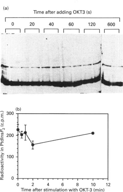
We have raised a panel of monoclonal antibodies to PtdIns4P 5-kinase C purified from bovine brain [Divecha, Brooksbank and Irvine (1992) Biochem. J. 288, 637-642]. This panel includes antibodies which specifically recognize PtdIns4P 5-kinase C both in a native catalytically active condition, and/or when presented on Western blots. Some of the former antibodies will also inhibit PtdIns4P 5-kinase C activity. We have used the blotting antibodies to study the bovine tissue distribution of PtdIns4P 5-kinase C and its distribution in mammalian species. We have also studied its localization in Jurkat cells and found it to be predominantly bound to membranes, with only a minority localized to the cytoskeleton. Neither PtdIns4P 5-kinase activity nor PtdIns4P 5-kinase C, as detected by Western blotting, were increased in the cytoskeleton after stimulation of Jurkat cells with OKT3. These antibodies should prove to be extremely useful tools with which to study the regulation of PtdIns4P 5-kinase C.
我们制备了一组针对从牛脑纯化的磷脂酰肌醇4-磷酸5-激酶C的单克隆抗体[迪韦查、布鲁克斯班克和欧文(1992年)《生物化学杂志》288卷,637 - 642页]。该组抗体包括能在天然催化活性状态下以及/或者在蛋白质印迹中特异性识别磷脂酰肌醇4-磷酸5-激酶C的抗体。部分前者抗体还会抑制磷脂酰肌醇4-磷酸5-激酶C的活性。我们已使用印迹抗体研究磷脂酰肌醇4-磷酸5-激酶C在牛组织中的分布及其在哺乳动物物种中的分布。我们还研究了其在Jurkat细胞中的定位,发现它主要与膜结合,只有少数定位于细胞骨架。在用OKT3刺激Jurkat细胞后,细胞骨架中通过蛋白质印迹检测到的磷脂酰肌醇4-磷酸5-激酶活性和磷脂酰肌醇4-磷酸5-激酶C均未增加。这些抗体应被证明是研究磷脂酰肌醇4-磷酸5-激酶C调节的极为有用的工具。