Malek A M, Gibbons G H, Dzau V J, Izumo S
Harvard Medical School-Massachusetts Institute of Technology, Division of Health Sciences and Technology, Boston.
J Clin Invest. 1993 Oct;92(4):2013-21. doi: 10.1172/JCI116796.
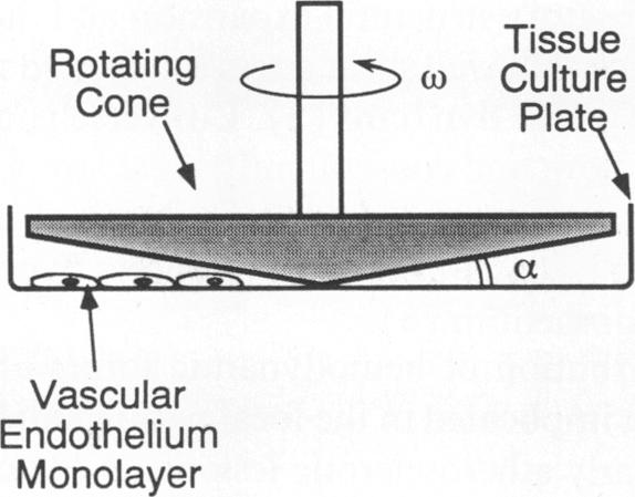
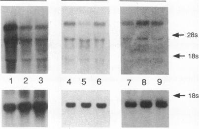
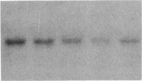
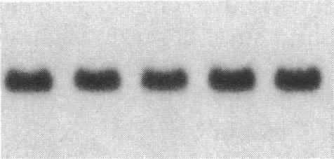
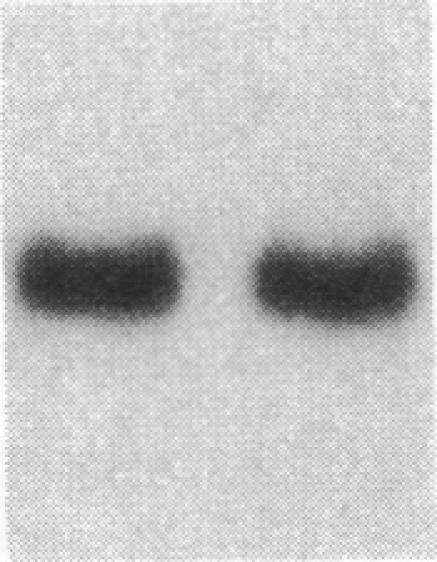
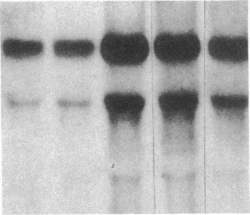
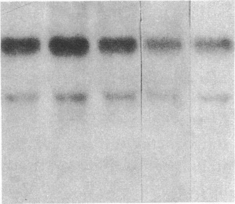
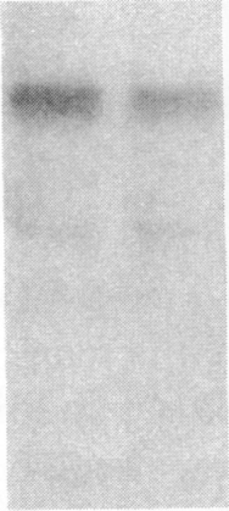
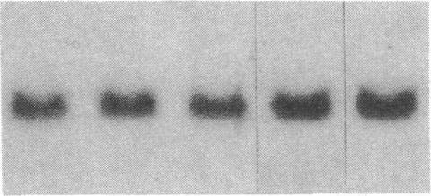
Fluid shear stress has been shown to be an important regulator of vascular structure and function through its effect on the endothelial cell. We have explored the effect of shear stress on the expression of the heparin-binding growth factors platelet-derived growth factor B chain (PDGF-B) and basic fibroblast growth factor (bFGF) in bovine aortic endothelial cells using a purpose-built cone-plate viscometer. Using morphometric analysis, we have mimicked the endothelial cell shape changes encountered in vivo in response to shear stress and correlated these with changes in gene expression. Steady laminar shear stress of 15 and 36 dyn/cm2 both resulted in endothelial cell shape change, but the higher shear stress induced greater and more uniform alignment in the direction of flow and nuclear protrusion after 24 h. Steady laminar shear stress of both 15 and 36 dyn/cm2 induced a significant 3.9- and 4.2-fold decrease, respectively, in PDGF-B mRNA at 9 h. In contrast, steady laminar shear of 15 dyn/cm2 induced a mild and transient 1.5-fold increase in bFGF mRNA while shear of 36 dyn/cm2 induced a significant 4.8-fold increase at 6 h of shear which remained at 2.9-fold at 9 h. Pulsatile and turbulent shear stress showed the same effect as steady laminar shear stress (all at 15 dyn/cm2 time-average magnitude) on PDGF-B and bFGF mRNA content. Cyclic stretch (20% strain, 20/min) of cells grown on silicone substrate did not significantly affect either PDGF-B or bFGF mRNA levels. These results suggest that expression of each peptide growth factor gene is differentially regulated by fluid shear stress in the vascular endothelial cell. These results may have implications on vascular structure and function in response to hemodynamic forces and present a model for the study of transduction of mechanical stimuli into altered gene expression.
流体剪切应力已被证明是通过对内皮细胞的作用来调节血管结构和功能的重要因素。我们使用特制的锥板粘度计,研究了剪切应力对牛主动脉内皮细胞中肝素结合生长因子血小板衍生生长因子B链(PDGF-B)和碱性成纤维细胞生长因子(bFGF)表达的影响。通过形态计量分析,我们模拟了体内内皮细胞在剪切应力作用下的形状变化,并将其与基因表达的变化相关联。15和36 dyn/cm2的稳定层流剪切应力均导致内皮细胞形状改变,但较高的剪切应力在24小时后诱导细胞在流动方向上更大且更均匀的排列以及核突出。15和36 dyn/cm2的稳定层流剪切应力在9小时时分别导致PDGF-B mRNA显著下降3.9倍和4.2倍。相比之下,15 dyn/cm2的稳定层流剪切在6小时时诱导bFGF mRNA轻度短暂增加1.5倍,而36 dyn/cm2的剪切在6小时时诱导显著增加4.8倍,在9小时时仍为2.9倍。脉动和湍流剪切应力对PDGF-B和bFGF mRNA含量的影响与稳定层流剪切应力(均为15 dyn/cm2时间平均幅度)相同。在硅胶基质上生长的细胞进行循环拉伸(20%应变,20次/分钟)对PDGF-B或bFGF mRNA水平均无显著影响。这些结果表明,血管内皮细胞中每种肽生长因子基因的表达受到流体剪切应力的差异调节。这些结果可能对血管结构和功能对血流动力学力的反应具有启示意义,并为研究机械刺激转化为基因表达改变提供了一个模型。