Okabayashi M, Angell M G, Budgeon L R, Kreider J W
Department of Pathology, Milton S. Hershey Medical Center, Pennsylvania State University, Hershey 17033.
Am J Pathol. 1993 Feb;142(2):489-96.
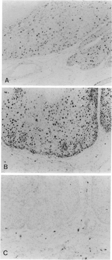
Lesions generated by infection with cottontail rabbit papillomavirus frequently undergo spontaneous regression. The purpose of this immunohistochemical study was to compare leukocyte and papilloma cell proliferation in progressing and regressing papillomas and to test the hypothesis that regression was associated with an inhibition of papilloma cell proliferation. The monoclonal antibodies (MAbs) MAb-019 (specific for DNA/bromodeoxyuridine [BrdU] complexes), Ki-67 (specific for actively proliferating cells), L11/135 (specific for rabbit T cells), and 2C4 (specific for rabbit class II antigen) were used for this purpose. In progressing papillomas, there were few leukocytes (< 1%) in the dermis that stained with MAb-019 and Ki-67, whereas these antibodies stained 4.5% and 6.8% of the intraepidermal leukocytes, respectively. Regressing papillomas contained conspicuous leukocytic infiltrates in the dermis, of which 76.9% were L11/135-positive T cells. However, few intradermal leukocytes (< 3%) stained positively with MAb-019 and Ki-67 MAbs, despite expressing rabbit class II antigen. The epidermis of regressing papillomas contained a higher percentage of MAb-019- and Ki-67-positive leukocytes than the epidermis of progressing papillomas. Intraepidermal leukocytes in progressing and regressing papillomas consisted mainly of T cells stained by L11/135. It appeared that many dermal leukocytes (mainly T cells) form a non-cycling T cell population in both progressing and regressing papillomas, whereas intraepidermal T cells in regressing papillomas were effectively activated and represented a cycling T cell population. MAb-019 and Ki-67 MAbs demonstrated similar staining patterns in papilloma and normal tissues. However, in both progressing and regressing papillomas, the Ki-67 MAb usually stained a larger percentage of cells than the MAb-019 MAb. MAb-019 and Ki-67 MAbs showed a homogeneous distribution of positive cells from basal layer to the upper layer in progressing papillomas. On the other hand, in regressing papillomas, cell staining with the two antibodies was concentrated in the basal and lower layers, but not in the upper layers. This result indicates that cell proliferation in the upper epidermal layers is suppressed in regressing papillomas. Our present data show that intraepidermal T- cell activation and suppression of tumor proliferation might play a crucial role in papilloma regression.
感染棉尾兔乳头瘤病毒所产生的病变常常会自发消退。本免疫组织化学研究的目的是比较进展期和消退期乳头瘤中白细胞和乳头瘤细胞的增殖情况,并验证消退与乳头瘤细胞增殖受抑制相关这一假说。为此使用了单克隆抗体(MAb):MAb - 019(对DNA/溴脱氧尿苷[BrdU]复合物具有特异性)、Ki - 67(对活跃增殖细胞具有特异性)、L11/135(对兔T细胞具有特异性)以及2C4(对兔II类抗原具有特异性)。在进展期乳头瘤中,真皮内用MAb - 019和Ki - 67染色的白细胞很少(<1%),而这些抗体分别对表皮内4.5%和6.8%的白细胞进行了染色。消退期乳头瘤在真皮内有明显的白细胞浸润,其中76.9%为L11/135阳性T细胞。然而,尽管真皮内白细胞表达兔II类抗原,但很少有细胞(<3%)用MAb - 019和Ki - 67单克隆抗体呈阳性染色。消退期乳头瘤的表皮中MAb - 019和Ki - 67阳性白细胞的比例高于进展期乳头瘤的表皮。进展期和消退期乳头瘤的表皮内白细胞主要由L11/135染色的T细胞组成。似乎在进展期和消退期乳头瘤中,许多真皮白细胞(主要是T细胞)形成了一个非循环T细胞群体,而消退期乳头瘤中的表皮内T细胞被有效激活,代表了一个循环T细胞群体。MAb - 019和Ki - 67单克隆抗体在乳头瘤组织和正常组织中表现出相似的染色模式。然而,在进展期和消退期乳头瘤中,Ki - 67单克隆抗体通常比MAb - 019单克隆抗体染色的细胞比例更高。在进展期乳头瘤中,MAb - 019和Ki - 67单克隆抗体显示阳性细胞从基底层到上层呈均匀分布。另一方面,在消退期乳头瘤中,两种抗体的细胞染色集中在基底层和下层,而上层没有。这一结果表明消退期乳头瘤表皮上层的细胞增殖受到抑制。我们目前的数据表明,表皮内T细胞激活和肿瘤增殖抑制可能在乳头瘤消退中起关键作用。