Heinzen R A, Hayes S F, Peacock M G, Hackstadt T
Rocky Mountain Laboratories, National Institute of Allergy and Infectious Diseases, Hamilton, Montana 59840.
Infect Immun. 1993 May;61(5):1926-35. doi: 10.1128/iai.61.5.1926-1935.1993.
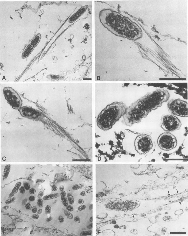
Members of the spotted fever group (SFG) of rickettsiae spread rapidly from cell to cell by an unknown mechanism(s). Staining of Rickettsia rickettsii-infected Vero cells with rhodamine phalloidin demonstrated unique actin filaments associated with one pole of intracellular rickettsiae. F-actin tails greater than 70 microns in length were seen extending from rickettsiae. Treatment of infected cells with chloramphenicol eliminated rickettsia-associated F-actin tails, suggesting that de novo protein synthesis of one or more rickettsial proteins is required for tail formation. Rickettsiae were coated with F-actin as early as 15 min postinfection, and tail formation was detected by 30 min. A survey of virulent and avirulent species within the SFG rickettsiae demonstrated that all formed actin tails. Typhus group rickettsiae, which do not spread directly from cell to cell, lacked F-actin tails entirely or exhibited only very short tails. Transmission electron microscopy demonstrated fibrillar material in close association with R. rickettsii but not Rickettsia prowazekii. Biochemical evidence that actin polymerization plays a role in movement was provided by showing that transit of R. rickettsii from infected cells into the cell culture medium was inhibited by treatment of host cells with cytochalasin D. These data suggest that the cell-to-cell transmission of SFG rickettsiae may be aided by induction of actin polymerization in a fashion similar to that described for Shigella flexneri and Listeria monocytogenes.
斑点热群立克次体通过未知机制在细胞间迅速传播。用罗丹明鬼笔环肽对感染立氏立克次体的非洲绿猴肾细胞进行染色,结果显示独特的肌动蛋白丝与细胞内立克次体的一极相关。观察到从立克次体延伸出长度超过70微米的丝状肌动蛋白尾。用氯霉素处理感染细胞可消除与立克次体相关的丝状肌动蛋白尾,这表明尾的形成需要一种或多种立克次体蛋白的从头合成。感染后15分钟,立克次体就被丝状肌动蛋白包被,30分钟时检测到尾的形成。对斑点热群立克次体中的强毒株和无毒株进行的一项调查表明,所有毒株都能形成肌动蛋白尾。不能在细胞间直接传播的斑疹伤寒群立克次体完全没有丝状肌动蛋白尾,或者仅表现出非常短的尾。透射电子显微镜显示,丝状物质与立氏立克次体紧密相关,但与普氏立克次体无关。用细胞松弛素D处理宿主细胞,可抑制立氏立克次体从感染细胞进入细胞培养基,这为肌动蛋白聚合在运动中起作用提供了生化证据。这些数据表明,斑点热群立克次体的细胞间传播可能以类似于福氏志贺菌和单核细胞增生李斯特菌的方式通过诱导肌动蛋白聚合来实现。