Wike D A, Tallent G, Peacock M G, Ormsbee R A
Infect Immun. 1972 May;5(5):715-22. doi: 10.1128/iai.5.5.715-722.1972.
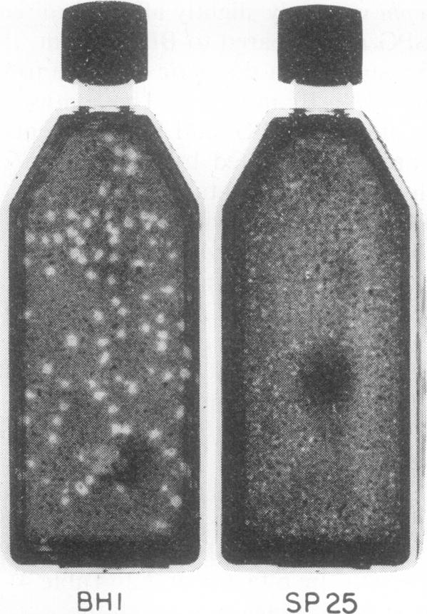
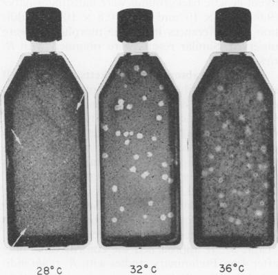
A plaque assay system for pathogenic rickettsiae, which utilizes primary chick embryo tissue cultures, is described. It proved to be a highly reproducible measure of infectiousness for Rickettsia rickettsi and R. typhi, which were employed in most studies; as well as for R. canada, R. prowazeki, R. sibirica, R. akari, R. conori, and Coxiella burneti. Plaque-forming units (PFU) were compared to direct rickettsial counts and to 50% infectious dose (ID(50)) values for embryonated eggs, mice, and guinea pigs. Plaque size, appearance, and number were influenced by diluent, incubation temperature after nutrient overlay, centrifugation of inoculated tissue cultures, and number of host cells planted initially in each flask. The most critical factors in plaque formation were diluent used in making rickettsial suspensions and incubation temperature (32 C) after nutrient overlay. Brain Heart Infusion was the only diluent capable of preventing significant delay in plaque formation and decreases in PFU and mouse ID(50). Plaque formation was unaffected by genetic background of host cells, volume of inoculum, temperature and length of incubation period before nutrient overlay, and rapid freezing and thawing of rickettsial seed. Centrifugation of inoculated cultures at 600 x g resulted in 100% irreversible absorption of rickettsiae to host cells within 5 min, whereas without centrifugation at least 4 hr was required to achieve the same effect.
本文描述了一种用于致病性立克次氏体的空斑测定系统,该系统利用原代鸡胚组织培养物。事实证明,对于大多数研究中使用的立氏立克次氏体和伤寒立克次氏体,以及加拿大立克次氏体、普氏立克次氏体、西伯利亚立克次氏体、小蛛立克次氏体、康氏立克次氏体和贝氏柯克斯体而言,它是一种高度可重复的感染性测量方法。将空斑形成单位(PFU)与立克次氏体直接计数以及对鸡胚、小鼠和豚鼠的50%感染剂量(ID50)值进行了比较。空斑大小、外观和数量受稀释剂、营养覆盖后的孵育温度、接种的组织培养物的离心以及每个培养瓶中最初接种的宿主细胞数量的影响。空斑形成的最关键因素是制备立克次氏体悬液时使用的稀释剂以及营养覆盖后的孵育温度(32℃)。脑心浸液是唯一能够防止空斑形成显著延迟以及PFU和小鼠ID50降低的稀释剂。空斑形成不受宿主细胞的遗传背景、接种物体积、营养覆盖前的孵育温度和时间以及立克次氏体种子的快速冻融的影响。对接种的培养物以600×g进行离心可使立克次氏体在5分钟内100%不可逆地吸附到宿主细胞上,而不进行离心则至少需要4小时才能达到相同效果。