Flint A J, Gebbink M F, Franza B R, Hill D E, Tonks N K
Cold Spring Harbor Laboratory, NY 11724.
EMBO J. 1993 May;12(5):1937-46. doi: 10.1002/j.1460-2075.1993.tb05843.x.
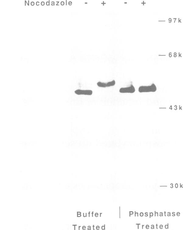
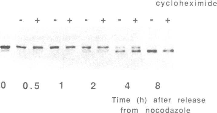
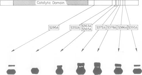
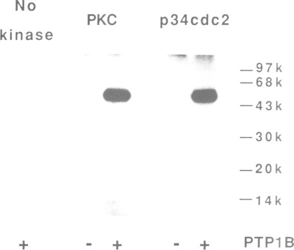
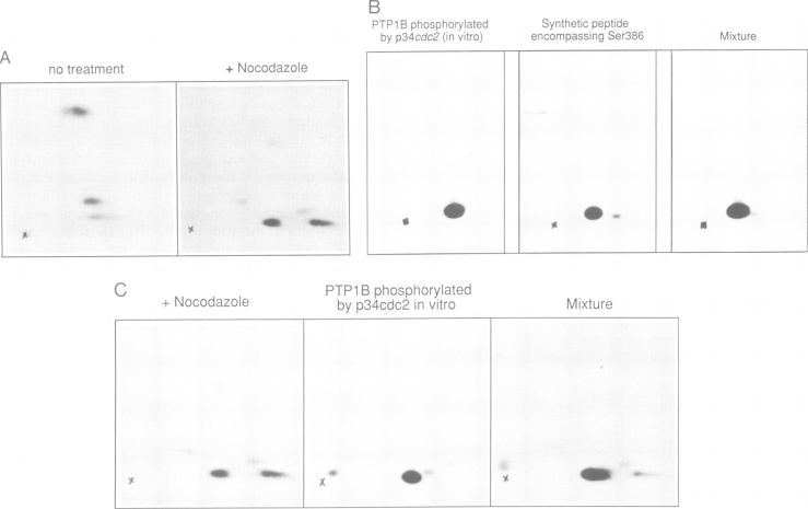
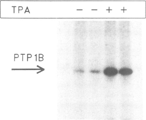
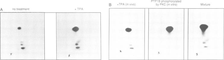
The non-transmembrane protein tyrosine phosphatase, PTP1B, comprises 435 amino acids, of which the C-terminal 114 residues have been implicated in controlling both localization and function of this enzyme. Inspection of the sequence of the C-terminal segment reveals a number of potential sites of phosphorylation. We show that PTP1B is phosphorylated on seryl residues in vivo. Increased phosphorylation of PTP1B is seen to accompany the transition from G2 to M phase of the cell cycle. Two major tryptic phosphopeptides appear in two-dimensional maps of PTP1B from mitotic cells. One of these comigrates with the peptide generated following phosphorylation of PTP1B in vitro at Ser386 by the mitotic protein Ser/Thr kinase p34cdc2:cyclin B. The site of phosphorylation that is responsible for the pronounced retardation in the electrophoretic mobility of PTP1B from mitotic cells has been identified by site directed mutagenesis as Ser352. The identify of the kinase responsible for this modification is presently unknown. We also show that stimulation of HeLa cells with the phorbol ester TPA enhances phosphorylation of PTP1B. Two dimensional phosphopeptide mapping reveals that the bulk of the phosphate is in a single tryptic peptide. The site, identified as Ser378, is also the site of phosphorylation by protein kinase C (PKC) in vitro. Thus the TPA-stimulated phosphorylation of PTP1B in vivo appears to result directly from phosphorylation by PKC. The effect of phosphorylation on the activity of PTP1B has been examined in immunoprecipitates from TPA-treated and nocodazole-arrested cells. TPA treatment does not appear to affect activity directly, whereas the activity of PTP1B from nocodazole-arrested cells is only 70% of that from asynchronous populations.
非跨膜蛋白酪氨酸磷酸酶PTP1B由435个氨基酸组成,其中C端的114个残基与该酶的定位和功能控制有关。对C端片段序列的检查揭示了一些潜在的磷酸化位点。我们发现PTP1B在体内丝氨酸残基上发生磷酸化。在细胞周期从G2期向M期转变时,PTP1B的磷酸化增加。在有丝分裂细胞的PTP1B二维图谱中出现了两个主要的胰蛋白酶磷酸肽。其中一个与有丝分裂蛋白丝氨酸/苏氨酸激酶p34cdc2:细胞周期蛋白B在体外将PTP1B的Ser386磷酸化后产生的肽迁移一致。通过定点诱变确定,导致有丝分裂细胞中PTP1B电泳迁移率显著减慢的磷酸化位点是Ser352。目前尚不清楚负责这种修饰的激酶的身份。我们还发现,用佛波酯TPA刺激HeLa细胞可增强PTP1B的磷酸化。二维磷酸肽图谱显示,大部分磷酸位于单个胰蛋白酶肽中。该位点被确定为Ser378,也是蛋白激酶C(PKC)在体外进行磷酸化的位点。因此,TPA在体内刺激PTP1B的磷酸化似乎直接源于PKC的磷酸化。已在TPA处理和诺考达唑阻滞细胞的免疫沉淀中检测了磷酸化对PTP1B活性的影响。TPA处理似乎不会直接影响活性,而诺考达唑阻滞细胞中PTP1B的活性仅为异步群体中PTP1B活性的70%。