Whiteside C I, Cameron R, Munk S, Levy J
Department of Medicine, University of Toronto, Ontario, Canada.
Am J Pathol. 1993 May;142(5):1641-53.
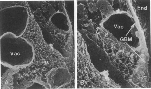
Puromycin aminonucleoside--(PAN) treated rats develop acute nephrotic syndrome, mimicking human minimal lesion disease. In PAN nephrosis, podocyte detachment from the glomerular basement membrane (GBM) is the most likely cause of massive proteinuria in this model. To elucidate further the mechanisms of PAN-induced cellular dysfunction, new methods were employed to visualize podocyte cytoskeletal aggregation and to measure fibrillar attachment to the GBM. Adult Sprague-Dawley rats (n = 4/group) received a single tail-vein injection of PAN (75 mg/kg). On days 1, 2, 3, and 5 following injection, 24-hour urine collections were obtained for creatinine clearance, albuminuria, and total proteinuria. Then kidneys from each group were fixed by perfusion. Podocytic cytoskeleton was visualized by scanning electron microscopy. Subepithelial GBM staining and attachment fiber number, observed on digitized images of transmission electron micrographs, were quantitated with computer-based density analysis. A significant reduction in attachment fiber number in the GBM lamina rara externa occurred by day 5. On scanning electron micrographs, the secondary and tertiary podocytic processes were observed to be formed by highly aggregated cytoskeleton, which became partially disaggregated by day 3, was totally absent by day 5, and normalized by day 20. Immunogold staining revealed that actin and vinculin localized to the tertiary podocytic processes in the normal state were dispersed into the cell body following PAN. Podocyte cytoskeletal disaggregation precedes, and detachment from the GBM occurs simultaneously with, the onset of massive proteinuria in the PAN model.
嘌呤霉素氨基核苷(PAN)处理的大鼠会发展为急性肾病综合征,类似于人类的微小病变疾病。在PAN肾病中,足细胞从肾小球基底膜(GBM)脱离是该模型中大量蛋白尿最可能的原因。为了进一步阐明PAN诱导细胞功能障碍的机制,采用了新的方法来观察足细胞细胞骨架聚集情况并测量纤维与GBM的附着情况。成年Sprague-Dawley大鼠(每组n = 4)通过尾静脉单次注射PAN(75 mg/kg)。在注射后的第1、2、3和5天,收集24小时尿液以测定肌酐清除率、蛋白尿和总蛋白尿。然后每组的肾脏通过灌注固定。通过扫描电子显微镜观察足细胞细胞骨架。在透射电子显微镜数字化图像上观察到的上皮下GBM染色和附着纤维数量,通过基于计算机的密度分析进行定量。到第5天时,GBM外疏松层中的附着纤维数量显著减少。在扫描电子显微镜下,观察到次级和三级足细胞突起由高度聚集化的细胞骨架形成,到第3天时部分解聚,第5天时完全消失,第20天时恢复正常。免疫金染色显示,正常状态下定位于三级足细胞突起的肌动蛋白和纽蛋白在PAN处理后分散到细胞体中。在PAN模型中,足细胞细胞骨架解聚先于大量蛋白尿的发生,且与足细胞从GBM脱离同时出现。