Sato K, Nishikawa S, Nakano A
Department of Biological Sciences, Graduate School of Science, University of Tokyo, Japan.
Mol Biol Cell. 1995 Nov;6(11):1459-77. doi: 10.1091/mbc.6.11.1459.
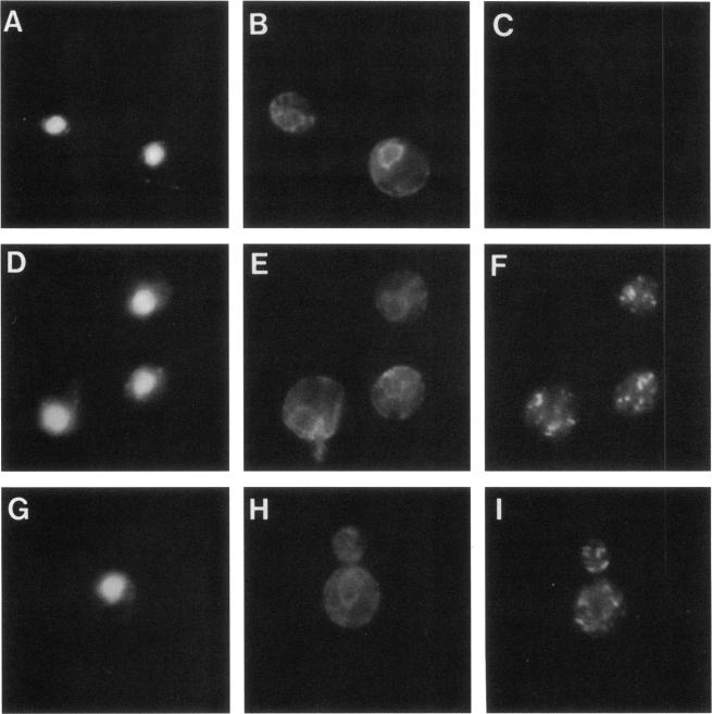
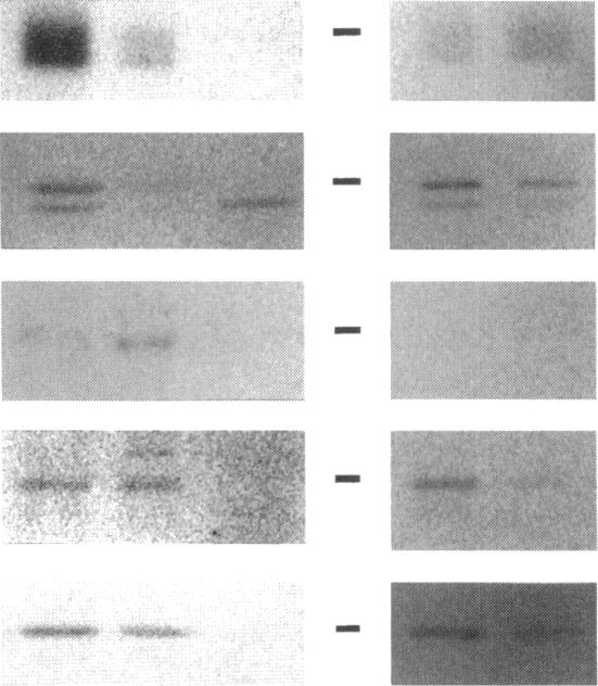
Yeast Sec12p, a type II transmembrane glycoprotein, is required for formation of transport vesicles from the endoplasmic reticulum (ER). Biochemical and morphological analyses have suggested that Sec12p is localized to the ER by two mechanisms: static retention in the ER and dynamic retrieval from the early region of the Golgi apparatus. The rer1 mutant we isolated in a previous study mislocalizes the authentic Sec12p to the later compartments of the Golgi. To understand the role of RER1 on Sec12p localization, we cloned the gene and determined its reading frame. RER1 encodes a hydrophobic protein of 188 amino acid residues containing four putative membrane spanning domains. The rer1 null mutant is viable. Even in the rer1 disrupted cells, immunofluorescence of Sec12p stains the ER, implying that the retention system is still operating in the mutant. To determine the subcellular localization of Rer1p, an epitope derived from the influenza hemagglutinin was added to the C-terminus of Rer1p and the cells expressing this tagged but functional protein were observed by immunofluorescence microscopy. The anti-HA monoclonal antibody stains the cells in a punctate pattern that is typical for Golgi proteins and clearly distinct from the ER staining. This punctate staining was in fact exaggerated in the sec7 mutant that accumulates the Golgi membranes at the restrictive temperature. Furthermore, double staining of Rer1p and Ypt1p, a GTPase that is known to reside in the Golgi apparatus, showed good colocalization. Subcellular fractionation experiments indicated that the fractionation pattern of Rer1p was similar to that of an early Golgi protein, Och1p. From these results, we suggest that Rer1p functions in the Golgi membrane to return Sec12p that has escaped from the static retention system of the ER.
酵母Sec12p是一种II型跨膜糖蛋白,是内质网(ER)形成运输小泡所必需的。生化和形态学分析表明,Sec12p通过两种机制定位于内质网:在内质网中的静态保留和从高尔基体早期区域的动态回收。我们在先前的研究中分离出的rer1突变体将真正的Sec12p错误定位于高尔基体的后期区室。为了了解RER1在Sec12p定位中的作用,我们克隆了该基因并确定了其阅读框。RER1编码一个由188个氨基酸残基组成的疏水蛋白,含有四个假定的跨膜结构域。rer1缺失突变体是有活力的。即使在rer1缺失的细胞中,Sec12p的免疫荧光也能对内质网进行染色,这意味着突变体中的保留系统仍在运行。为了确定Rer1p的亚细胞定位,将来自流感血凝素的表位添加到Rer1p的C末端,并通过免疫荧光显微镜观察表达这种带有标签但功能正常蛋白质的细胞。抗HA单克隆抗体以点状模式对细胞进行染色,这是高尔基体蛋白的典型模式,与内质网染色明显不同。事实上,这种点状染色在sec7突变体中被放大,该突变体在限制温度下积累高尔基体膜。此外,Rer1p和Ypt1p(一种已知存在于高尔基体中的GTP酶)的双重染色显示出良好的共定位。亚细胞分级分离实验表明,Rer1p的分级分离模式与早期高尔基体蛋白Och1p相似。从这些结果中,我们认为Rer1p在高尔基体膜中发挥作用,使从内质网静态保留系统中逃逸的Sec12p返回。