Nishikawa S, Hirata A, Nakano A
Department of Plant Sciences, Graduate School of Science, University of Tokyo, Japan.
Mol Biol Cell. 1994 Oct;5(10):1129-43. doi: 10.1091/mbc.5.10.1129.
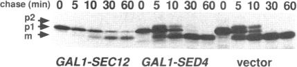
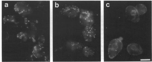
Immunofluorescence staining of yeast cells with anti-binding protein (BiP) antibodies shows uniform staining of the endoplasmic reticulum (ER). We have found that overproduction of Sec12p, an ER membrane protein, causes a change of BiP distribution within the cell. Upon induction of Sec12p by the GAL1 promoter, the staining pattern of BiP turns into bright dots scattering in the cell, whereas the staining of Sec12p remains to be the typical ER figure. Overproduction of other ER membrane proteins, HMG-CoA reductase or Sed4 protein, does not induce such relocalization of BiP. Pulse-chase experiments and electron microscopy have revealed that the overproduction of Sec12p inhibits protein transport from the ER to the Golgi apparatus. When the transport is arrested by one of the sec mutations that block the ER-to-Golgi step at the restrictive temperature, the BiP staining also changes into the punctate pattern. In contrast, the sec mutants that block later or earlier steps of the secretory pathway do not induce such change of BiP localization. These observations indicate that relocalization of BiP is caused by the inhibition of ER-to-Golgi transport. Using immunoelectron microscopy, we have found that the punctate staining is because of the accumulation of BiP in the restricted region of the ER, which we propose to call the "BiP body." This implicates existence of ER subdomains in yeast. A vacuolar protein, proteinase A, appears to colocalize in the BiP body when the ER-to-Golgi transport is blocked, suggesting that the BiP body may have a role as the site of accumulation of cargo molecules before exit from the ER.
用抗结合蛋白(BiP)抗体对酵母细胞进行免疫荧光染色,结果显示内质网(ER)呈现均匀染色。我们发现,内质网膜蛋白Sec12p的过量表达会导致BiP在细胞内的分布发生变化。当通过GAL1启动子诱导Sec12p表达时,BiP的染色模式转变为在细胞内分散的亮点,而Sec12p的染色仍保持典型的内质网形态。其他内质网膜蛋白,如HMG-CoA还原酶或Sed4蛋白的过量表达,并不会诱导BiP发生这种重新定位。脉冲追踪实验和电子显微镜观察表明,Sec12p的过量表达会抑制蛋白质从内质网向高尔基体的转运。当在限制温度下,通过一个阻断内质网到高尔基体步骤的sec突变体使转运停滞时,BiP染色也会变为点状模式。相反,阻断分泌途径后期或早期步骤的sec突变体不会诱导BiP定位发生这种变化。这些观察结果表明,BiP的重新定位是由内质网到高尔基体转运的抑制引起的。通过免疫电子显微镜,我们发现点状染色是由于BiP在内质网的受限区域积累所致,我们将该区域称为“BiP体”。这暗示酵母中存在内质网亚结构域。当内质网到高尔基体的转运受阻时,一种液泡蛋白,蛋白酶A,似乎与BiP体共定位,这表明BiP体可能在货物分子从内质网输出之前作为其积累位点发挥作用。