Schultz P, Olland S, Oudet P, Hancock R
Institut de Génétique et de Biologie Moléculaire et Cellulaire, CentreNational de la Recherche Scientifique, Institut National de la Sante et de la Recherche Médicale, Université Louis Pasteur, Canada.
Proc Natl Acad Sci U S A. 1996 Jun 11;93(12):5936-40. doi: 10.1073/pnas.93.12.5936.
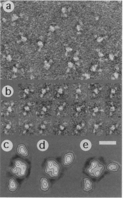
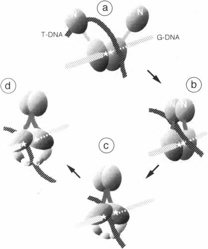
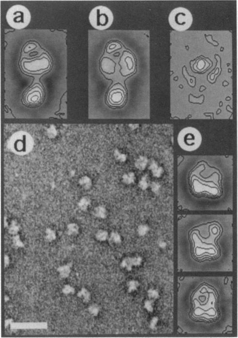
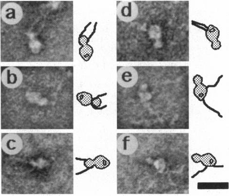
Type II DNA topoisomerases, which create a transient gate in duplex DNA and transfer a second duplex DNA through this gate, are essential for topological transformations of DNA in prokaryotic and eukaryotic cells and are of interest not only from a mechanistic perspective but also because they are targets of agents for anticancer and antimicrobial chemotherapy. Here we describe the structure of the molecule of human topoisomerase II [DNA topoisomerase (ATP-hydrolyzing), EC 5.99.1.3] as seen by scanning transmission electron microscopy. A globular approximately 90-angstrom diameter core is connected by linkers to two approximately 50-angstrom domains, which were shown by comparison with genetically truncated Saccharomyces cerevisiae topoisomerase II to contain the N-terminal region of the approximately 170-kDa subunits and that are seen in different orientations. When the ATP-binding site is occupied by a nonhydrolyzable ATP analog, a quite different structure is seen that results from a major conformational change and consists of two domains approximately 90 angstrom and approximately 60 angstrom in diameter connected by a linker, and in which the N-terminal domains have interacted. About two-thirds of the molecules show an approximately 25 A tunnel in the apical part of the large domain, and the remainder contain an internal cavity approximately 30 A wide in the large domain close to the linker region. We propose that structural rearrangements lead to this displacement of an internal tunnel. The tunnel is likely to represent the channel through which one DNA duplex, after capture in the clamp formed by the N-terminal domains, is transferred across the interface between the enzyme's subunits. These images are consistent with biochemical observations and provide a structural basis for understanding the reaction of topoisomerase II.
II型DNA拓扑异构酶可在双链DNA中形成一个瞬时门,并使另一条双链DNA通过此门,它对于原核细胞和真核细胞中DNA的拓扑转变至关重要,不仅从机制角度来看很有趣,还因为它们是抗癌和抗菌化疗药物的作用靶点。在此,我们描述了通过扫描透射电子显微镜观察到的人拓扑异构酶II分子[DNA拓扑异构酶(ATP水解),EC 5.99.1.3]的结构。一个直径约90埃的球状核心通过连接子与两个约50埃的结构域相连,通过与基因截短的酿酒酵母拓扑异构酶II比较表明,这两个结构域包含约170 kDa亚基的N端区域,且呈现出不同的取向。当ATP结合位点被一种不可水解的ATP类似物占据时,会观察到一种截然不同的结构,这是由主要的构象变化导致的,它由两个直径分别约为90埃和约60埃的结构域通过一个连接子相连组成,其中N端结构域相互作用。约三分之二的分子在大结构域的顶端部分显示出一个约25埃的通道,其余的在靠近连接子区域的大结构域中包含一个约30埃宽的内部腔。我们提出结构重排导致了这个内部通道的移位。这个通道可能代表了一条DNA双链在被N端结构域形成的夹子捕获后,穿过酶亚基之间界面的转移通道。这些图像与生化观察结果一致,并为理解拓扑异构酶II的反应提供了结构基础。