Luther T, Flössel C, Mackman N, Bierhaus A, Kasper M, Albrecht S, Sage E H, Iruela-Arispe L, Grossmann H, Ströhlein A, Zhang Y, Nawroth P P, Carmeliet P, Loskutoff D J, Müller M
Institute of Pathology, Technical University Dresden, Dresden, Germany.
Am J Pathol. 1996 Jul;149(1):101-13.
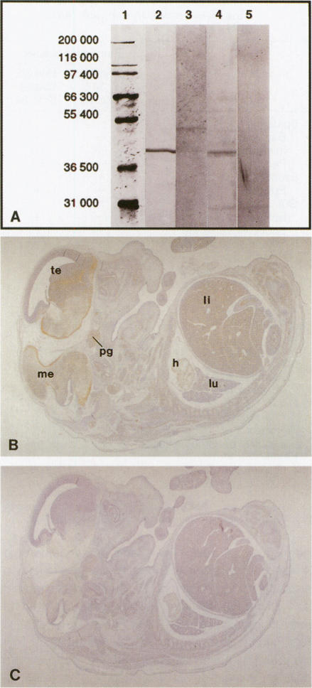
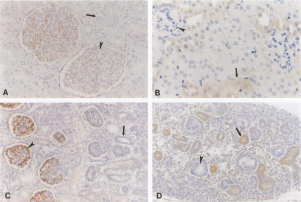
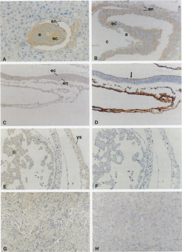
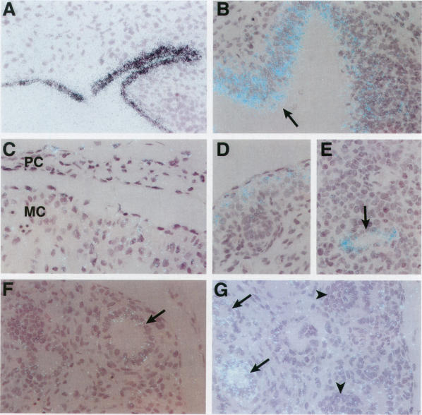
In the adult organism the cellular distribution of tissue factor (TF) expression corresponds to biological boundary layers forming a hemostatic barrier ready to activate blood coagulation after tissue injury. Whether TF expression might also play a role in development is unknown. To determine the significance of TF in ontogenesis, we examined the pattern of TF expression in mouse development and compared it with the distribution of TF in human post-implantation embryos and fetuses of corresponding gestational age. At early embryonic periods of murine (6.5 and 7.5 pc) and human (stage 5) development, there was strong expression of TF in both ectodermal and entodermal cells. In situ hybridization and immunohistochemistry demonstrated that TF mRNA and protein were expressed widely in epithelial areas with high levels of morphogenic activity during organogenesis. Staining for TF was seen during ontogenetic development in tissues such as epidermis, myocardium, bronchial epithelium, and hepatocytes, which express TF in the adult organism. Surprisingly, during renal development and in adults, expression of TF differed between humans and mice. In humans, maturing stage glomeruli were stained for TF whereas in mice, TF was absent from glomeruli but was present in the epithelia of tubular segments. In neuroepithelial cells, there was a substantial expression of TF. Moreover, there was robust TF expression in tissues such as skeletal muscle and pancreas, which do not express it in the adult. In contrast, expression of the physiological ligand for TF, factor VII, was not detectable during early stages of human embryogenesis using immunohistochemistry. The temporal and spatial pattern of TF expression during murine and human development supports the contention that TF serves as an important morphogenic factor during embryogenesis.
在成年生物体中,组织因子(TF)表达的细胞分布与形成止血屏障的生物边界层相对应,该屏障在组织损伤后可随时激活血液凝固。TF表达在发育过程中是否也起作用尚不清楚。为了确定TF在个体发生中的重要性,我们研究了小鼠发育过程中TF的表达模式,并将其与相应孕周的人类植入后胚胎和胎儿中TF的分布进行了比较。在小鼠(妊娠6.5和7.5天)和人类(第5阶段)发育的早期胚胎阶段,外胚层和内胚层细胞中均有强烈的TF表达。原位杂交和免疫组织化学表明,在器官发生过程中,TF mRNA和蛋白在具有高水平形态发生活性的上皮区域广泛表达。在个体发育过程中,在表皮、心肌、支气管上皮和肝细胞等组织中可见TF染色,这些组织在成年生物体中表达TF。令人惊讶的是,在肾脏发育过程中和成年期,人类和小鼠之间TF的表达存在差异。在人类中,成熟阶段的肾小球有TF染色,而在小鼠中,肾小球中没有TF,但在肾小管节段的上皮细胞中有TF。在神经上皮细胞中,有大量的TF表达。此外,在骨骼肌和胰腺等组织中也有强烈的TF表达,而这些组织在成年期不表达TF。相反,使用免疫组织化学在人类胚胎发育早期未检测到TF的生理配体因子VII的表达。小鼠和人类发育过程中TF表达的时空模式支持了TF在胚胎发生过程中作为重要形态发生因子的观点。