Schlosser S F, Burgstahler A D, Nathanson M H
Liver Study Unit, Yale University School of Medicine, New Haven, CT 06520, USA.
Proc Natl Acad Sci U S A. 1996 Sep 3;93(18):9948-53. doi: 10.1073/pnas.93.18.9948.
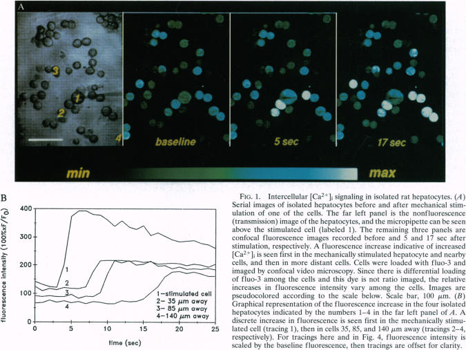
Intercellular communication among certain cell types can occur via ATP secretion, which leads to stimulation of nucleotide receptors on target cells. In epithelial cells, however, intercellular communication is thought to occur instead via gap junctions. Here we examined whether one epithelial cell type, hepatocytes, can also communicate via nucleotide secretion. The effects on cytosolic Ca2+ ([Ca2+]i) of mechanical stimulation, including microinjection, were examined in isolated rat hepatocytes and in isolated bile duct units using confocal fluorescence video microscopy. Mechanical stimulation of a single hepatocyte evoked an increase in [Ca2+]i in the stimulated cell plus an unexpected [Ca2+]i rise in neighboring noncontacting hepatocytes. Perifusion with ATP before mechanical stimulation suppressed the [Ca2+]i increase, but pretreatment with phenylephrine did not. The P2 receptor antagonist suramin inhibited these intercellular [Ca2+]i signals. The ATP/ADPase apyrase reversibly inhibited the [Ca2+]i rise induced by mechanical stimulation, and did not block vasopressin-induced [Ca2+]i signals. Mechanical stimulation of hepatocytes also induced a [Ca2+]i increase in cocultured isolated bile duct units, and this [Ca2+]i increase was inhibited by apyrase as well. Finally, this form of [Ca2+]i signaling could be elicited in the presence of propidium iodide without nuclear labeling by that dye, indicating that this phenomenon does not depend on disruption of the stimulated cell. Thus, mechanical stimulation of isolated hepatocytes, including by microinjection, can evoke [Ca2+]i signals in the stimulated cell as well as in neighboring noncontacting hepatocytes and bile duct epithelia. This signaling is mediated by release of ATP or other nucleotides into the extracellular space. This is an important technical consideration given the widespread use of microinjection techniques for examining mechanisms of signal transduction. Moreover, the evidence provided suggests a novel paracrine signaling pathway for epithelia, which previously were thought to communicate exclusively via gap junctions.
某些细胞类型之间的细胞间通讯可通过ATP分泌发生,这会导致靶细胞上的核苷酸受体受到刺激。然而,在上皮细胞中,细胞间通讯被认为是通过缝隙连接发生的。在这里,我们研究了一种上皮细胞类型——肝细胞是否也能通过核苷酸分泌进行通讯。我们使用共聚焦荧光视频显微镜,在分离的大鼠肝细胞和分离的胆管单位中,研究了包括显微注射在内的机械刺激对细胞溶质Ca2+([Ca2+]i)的影响。对单个肝细胞的机械刺激会导致受刺激细胞内的[Ca2+]i增加,以及相邻未接触肝细胞中意外的[Ca2+]i升高。在机械刺激前用ATP进行灌流可抑制[Ca2+]i的增加,但用去氧肾上腺素预处理则无效。P2受体拮抗剂苏拉明可抑制这些细胞间的[Ca2+]i信号。ATP/ADP酶(腺苷三磷酸双磷酸酶)可可逆地抑制机械刺激诱导的[Ca2+]i升高,且不阻断血管加压素诱导的[Ca2+]i信号。对肝细胞的机械刺激也会导致共培养的分离胆管单位中[Ca2+]i增加,且这种[Ca2+]i增加也会被腺苷三磷酸双磷酸酶抑制。最后,在存在碘化丙啶的情况下,这种形式的[Ca2+]i信号传导可以在没有该染料进行核标记的情况下引发,这表明这种现象不依赖于受刺激细胞的破坏。因此,对分离的肝细胞进行机械刺激,包括显微注射,可在受刺激细胞以及相邻未接触的肝细胞和胆管上皮细胞中引发[Ca2+]i信号。这种信号传导是由ATP或其他核苷酸释放到细胞外空间介导的。鉴于显微注射技术在研究信号转导机制中的广泛应用,这是一个重要的技术考量因素。此外,所提供的证据表明上皮细胞存在一种新的旁分泌信号通路,而此前认为上皮细胞仅通过缝隙连接进行通讯。