Emans N, Verkman A S
Department of Medicine, University of California, San Francisco 94143-0521 USA.
Biophys J. 1996 Jul;71(1):487-94. doi: 10.1016/S0006-3495(96)79250-0.
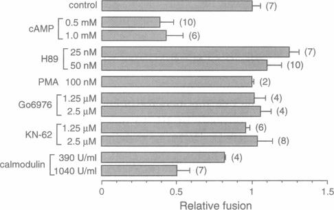
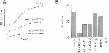
A quantitative real-time assay of cell-free endosomal vesicle fusion was developed and applied to study fusion mechanisms in endosomes from baby hamster kidney (BHK-21) cells. The assay is based on an irreversible approximately 10-fold increase in BODIPY-avidin fluorescence on binding of biotinylated conjugates. BODIPY-avidin and biotin-dextran were internalized for 10 min at 37 degrees C into separate populations of BHK-21 cells, and endosome fractions were prepared. Postnuclear supernatant fractions underwent ATP- and temperature-dependent fusion, as measured in a sensitive custom-built microfluorimeter by the continuous increase in BODIPY-avidin fluorescence. Fusion processes of efficiency > 2.5% could be detected with 200-ms time resolution in sample volumes of 50 microL containing endosomes derived from approximately 4 x 10(4) cells. The fusion time course consisted of a distinct lag phase (up to 10 min) in which little fusion occurred, followed by an approximately exponential rise (t 1/2 10-30 min; fusion efficiency approximately 15%). The lag phase was reduced by preincubation of separate endosome fractions with ATP at 37 degrees C and by coincubation of endosomes at 22 degrees C before the assay, suggesting a rate-limiting step involving binding of a soluble protein to the endosome membrane. Endosome fusion was strongly inhibited by GTP gamma S, N-ethylmaleimide, and AIF4-. Endosome fusion was not affected by phorbol myristate acetate but was significantly inhibited by cAMP and bovine brain calmodulin. The results establish a sensitive real-time fluorescence assay to quantify the kinetics and extent of endosome fusion in a cell-free system and demonstrate regulation of early endosome fusion by cytosolic second messengers.
我们开发了一种用于无细胞内体囊泡融合的定量实时检测方法,并将其应用于研究幼仓鼠肾(BHK-21)细胞内体的融合机制。该检测方法基于生物素化缀合物结合时BODIPY-抗生物素蛋白荧光不可逆地增加约10倍。将BODIPY-抗生物素蛋白和生物素-葡聚糖在37℃内化10分钟,分别导入不同群体的BHK-21细胞中,然后制备内体组分。核后上清液组分发生ATP和温度依赖性融合,通过灵敏的定制微荧光计测量BODIPY-抗生物素蛋白荧光的持续增加来检测。在含有源自约4×10⁴个细胞的内体的50微升样品体积中,以200毫秒的时间分辨率可以检测到效率>2.5%的融合过程。融合时间进程包括一个明显的延迟期(长达10分钟),在此期间几乎没有融合发生,随后是近似指数上升(t 1/2为10 - 30分钟;融合效率约为15%)。通过在37℃下将单独的内体组分与ATP预孵育以及在检测前于22℃下将内体共同孵育,延迟期缩短,这表明存在一个限速步骤,涉及一种可溶性蛋白与内体膜的结合。内体融合受到GTPγS、N-乙基马来酰亚胺和AIF4⁻的强烈抑制。内体融合不受佛波酯肉豆蔻酸酯乙酸盐的影响,但受到cAMP和牛脑钙调蛋白的显著抑制。这些结果建立了一种灵敏的实时荧光检测方法,用于在无细胞系统中定量内体融合的动力学和程度,并证明了胞质第二信使对早期内体融合的调节作用。