Ma N, Hunt N H, Madigan M C, Chan-Ling T
Department of Pathology, University of Sydney, Australia.
Am J Pathol. 1996 Nov;149(5):1745-62.
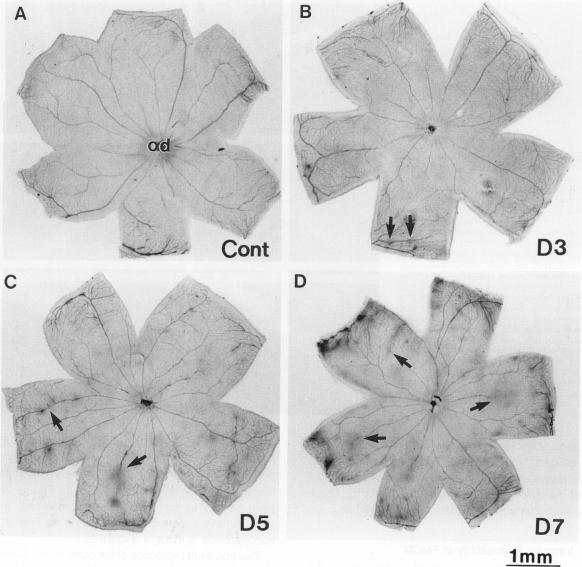
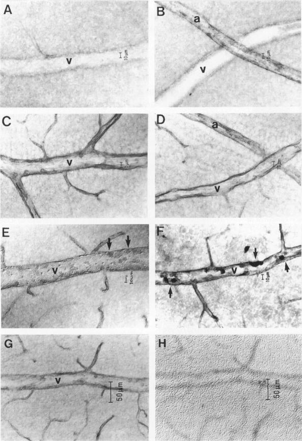
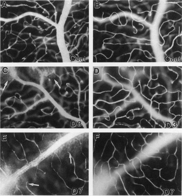
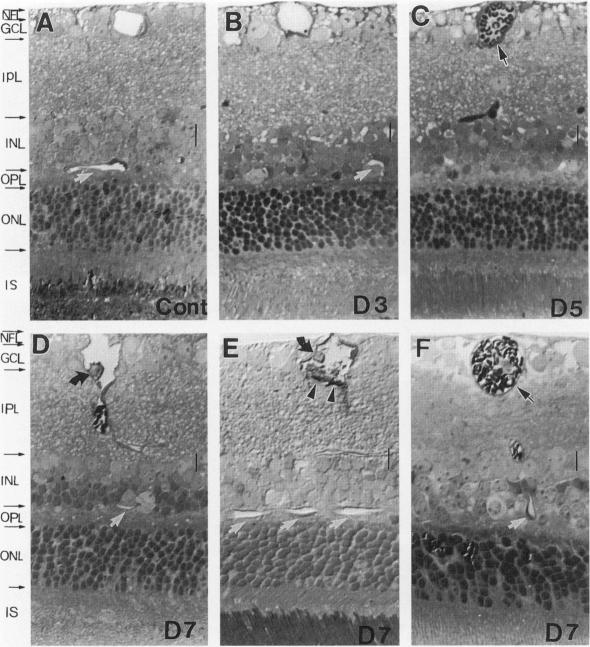
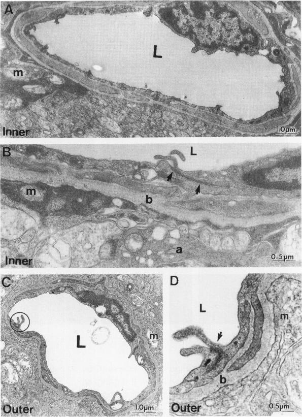
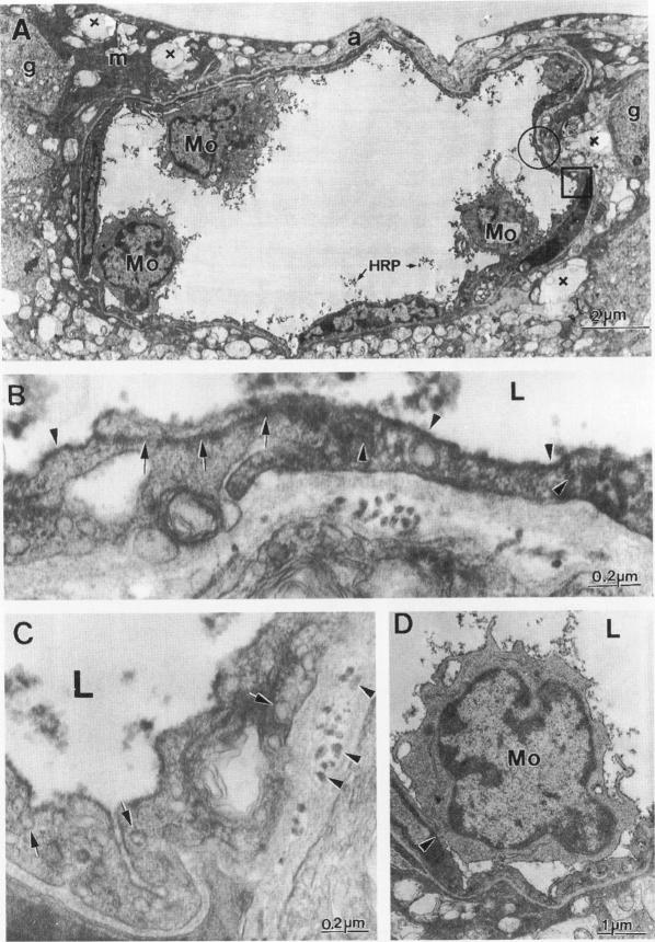
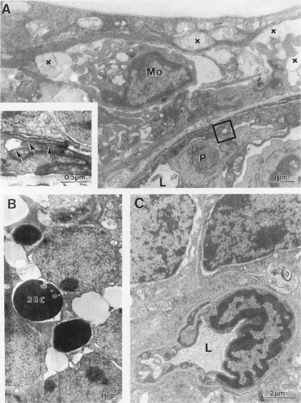
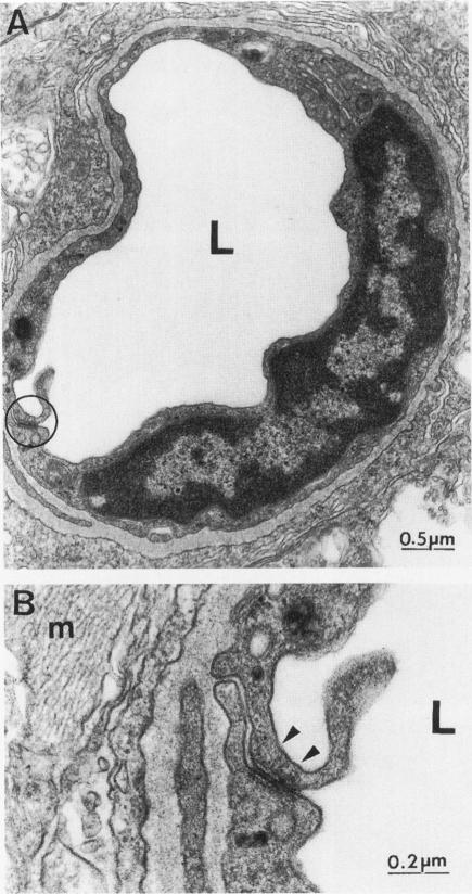
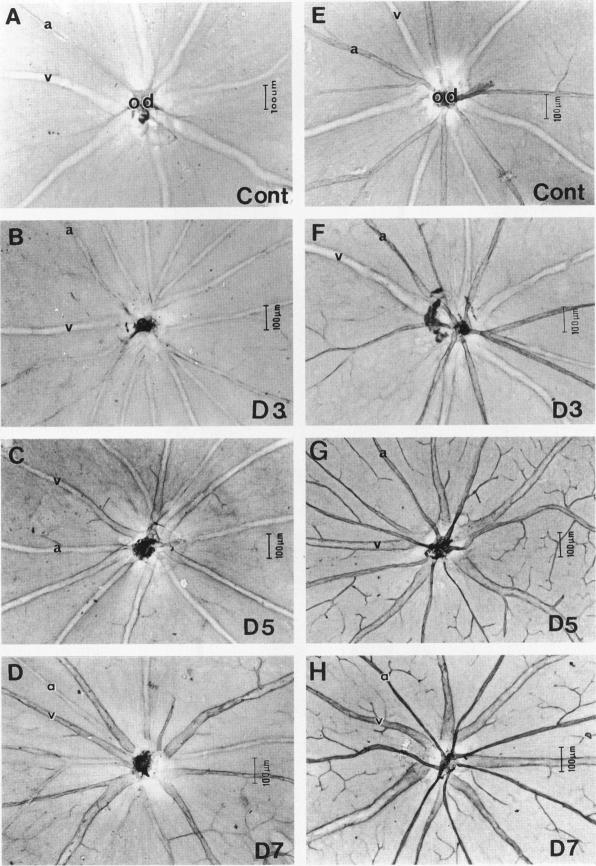
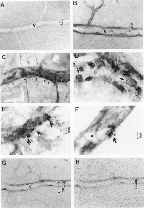
The relationships between increased vascular permeability to protein, monocyte adherence to the endothelium, and expression of the cell adhesion molecules, intercellular adhesion molecule-1 (ICAM-1) and vascular cell adhesion molecule-1 (VCAM-1) in the central nervous system microvasculature were studied during the progression of fatal murine cerebral malaria. CBA mice were inoculated with Plasmodium berghei ANKA, and changes in the retinal microvasculature were examined on days 3, 5, and 7 postinoculation (p.i.). Evans blue dye and horseradish peroxidase (HRP) were administered intravenously to assess vascular permeability to macromolecules macroscopically and by light and electron microscopy. ICAM-1 and VCAM-1 expression were examined by immunohistochemistry. HRP leakage into the retinal parenchyma was seen macroscopically at a low level on day 3 p.i., increasing progressively at day 5 (the earliest time at which cerebral symptoms were observed) and day 7 (the day on which animals showed severe behavioral abnormalities and died). The inner retinal vascular plexus showed a slight increase in vascular permeability to intravenous Evans blue at day 3 p.i. and congestion, monocyte adherence to the endothelium, and increased vascular permeability to both Evans blue and HRP at day 7 p.i. Electron microscopic observations were consistent with these findings and also revealed disrupted light junctions and the coating of monocytes and endothelium with HRP at day 7 p.i. Immunohistochemical staining and densitometry showed a progressive increase from day 3 to day 7 p.i. in the densities of ICAM-1 and VCAM-1 on the venular endothelium of the inner retinal vascular plexus, with the appearance of adherent ICAM-1+ monocytes at the terminal stage of the disease. None of the pathological changes associated with the inner retinal plexus were seen at any stage in the outer retinal plexus. These results suggest the following sequence of events in the inner retinal vessels, particularly the venules, during the progression of fatal murine cerebral malaria: 1) a mild increase in vascular permeability at approximately day 3 p.i., 2) a progressive increase in endothelial expression of the cell adhesion molecules ICAM-1 and VCAM-1, commencing at approximately day 3 p.i., 3) monocyte adhesion to the endothelium starting at approximately day 5 p.i., and 4) frank disruption of endothelial integrity at the terminal stage (day 7 p.i.), leading to edema and hemorrhage. Similar changes in cerebral vessels may underlie the neurological complications of the disease.
在致命性鼠脑型疟疾的病程中,研究了中枢神经系统微血管中蛋白质血管通透性增加、单核细胞黏附于内皮以及细胞黏附分子细胞间黏附分子-1(ICAM-1)和血管细胞黏附分子-1(VCAM-1)表达之间的关系。给CBA小鼠接种伯氏疟原虫ANKA株,并在接种后第3、5和7天检查视网膜微血管的变化。静脉注射伊文思蓝染料和辣根过氧化物酶(HRP),通过宏观观察以及光镜和电镜检查来评估大分子的血管通透性。通过免疫组织化学检查ICAM-1和VCAM-1的表达。接种后第3天,肉眼可见HRP少量渗漏到视网膜实质,在第5天(最早观察到脑部症状的时间)和第7天(动物出现严重行为异常并死亡的日子)逐渐增加。接种后第3天,视网膜内血管丛对静脉注射伊文思蓝的血管通透性略有增加,第7天出现充血、单核细胞黏附于内皮以及对伊文思蓝和HRP的血管通透性增加。电镜观察结果与这些发现一致,还显示在接种后第7天紧密连接被破坏,单核细胞和内皮被HRP覆盖。免疫组织化学染色和光密度测定显示,从接种后第3天到第7天,视网膜内血管丛小静脉内皮上ICAM-1和VCAM-1的密度逐渐增加,在疾病末期出现黏附的ICAM-1+单核细胞。在视网膜外血管丛的任何阶段均未观察到与视网膜内血管丛相关的病理变化。这些结果提示在致命性鼠脑型疟疾病程中,视网膜内血管尤其是小静脉中发生了以下一系列事件:1)接种后约第3天血管通透性轻度增加;2)从接种后约第3天开始,细胞黏附分子ICAM-1和VCAM-1在内皮细胞上的表达逐渐增加;3)接种后约第5天开始单核细胞黏附于内皮;4)疾病末期(接种后第7天)内皮完整性明显破坏,导致水肿和出血。脑血管的类似变化可能是该疾病神经并发症的基础。