Lin X H, Grako K A, Burg M A, Stallcup W B
La Jolla Cancer Research Center, Burnham Institute, California 92037, USA.
Mol Biol Cell. 1996 Dec;7(12):1977-93. doi: 10.1091/mbc.7.12.1977.
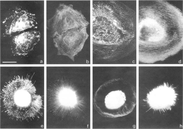
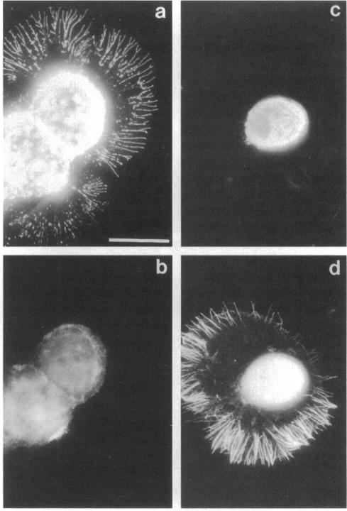
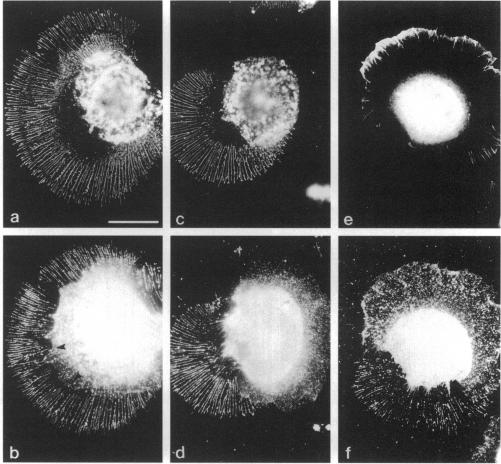
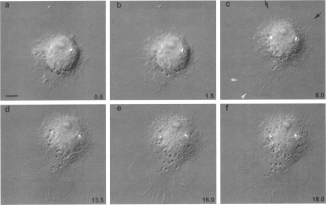
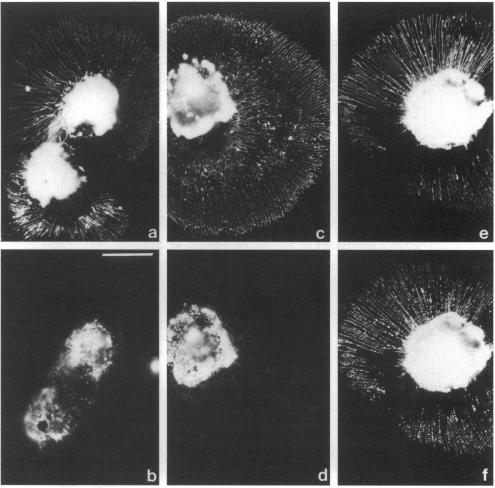
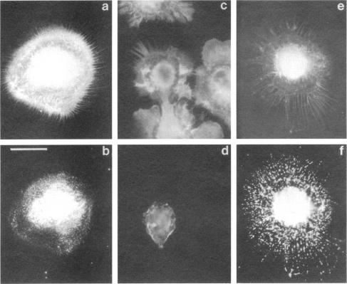
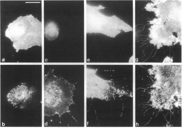
The transmembrane proteoglycan NG2 is able to interact both with components of the extracellular matrix and with the actin cytoskeleton. An examination of the distribution of NG2 during cell spreading suggests that NG2 can associate with two distinct types of actin-containing cytoskeletal structures, depending on the nature of the stimulus derived from the substratum. On fibronectin-coated dishes, cell surface NG2 associates exclusively with stress fibers developing within the cell. On poly-L-lysine-coated dishes, cell surface NG2 is associated with radial processes extending from the cell periphery. Spreading on fibronectin/poly-L-lysine mixtures, as well as on matrix components such as laminin, tenascin, and type VI collagen, produces cells with mosaic characteristics, i.e., NG2 is associated with both types of structures. NG2-positive radial processes are distinct from a second population of radial structures that contain fascin. NG2-positive extensions appear to be individual self-contained units (filopodia), whereas fascin is associated with actin ribs within sheets of membrane (lamellipodia). NG2- and fascin-positive structures are often localized to opposite poles of spreading cells, suggesting a possible role for the two classes of cellular extensions in the establishment of cell polarity during morphogenesis or migration. Time lapse imaging confirms the presence of lamellipodia on the leading edges of migrating cells, while numerous filopodia are present on trailing edges.
跨膜蛋白聚糖NG2既能与细胞外基质成分相互作用,也能与肌动蛋白细胞骨架相互作用。对细胞铺展过程中NG2分布的研究表明,根据源自基质的刺激性质,NG2可与两种不同类型的含肌动蛋白细胞骨架结构相关联。在纤连蛋白包被的培养皿上,细胞表面的NG2仅与细胞内形成的应力纤维相关联。在聚-L-赖氨酸包被的培养皿上,细胞表面的NG2与从细胞周边延伸的放射状突起相关联。在纤连蛋白/聚-L-赖氨酸混合物以及层粘连蛋白、腱生蛋白和VI型胶原等基质成分上铺展时,会产生具有镶嵌特征的细胞,即NG2与两种类型的结构都相关联。NG2阳性的放射状突起不同于含有成束蛋白的另一类放射状结构。NG2阳性的延伸似乎是独立的单元(丝状伪足),而成束蛋白与膜片(片状伪足)内的肌动蛋白肋相关联。NG2阳性和成束蛋白阳性的结构通常定位于铺展细胞的相对两极,这表明这两类细胞延伸在形态发生或迁移过程中细胞极性的建立中可能发挥作用。延时成像证实迁移细胞前缘存在片状伪足,而后缘存在大量丝状伪足。