Hussain S M, Stoker J, Zwamborn A W, Den Hollander J C, Kuiper J W, Entius C A, Laméris J S
Department of Radiology, Eramus University, Rotterdam, The Netherlands.
J Anat. 1996 Dec;189 ( Pt 3)(Pt 3):677-82.
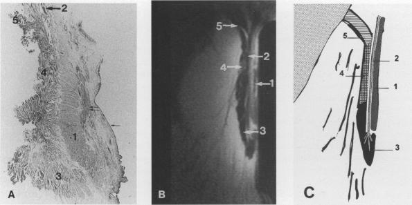
The purpose of this study was to correlate the in vivo endoanal MRI findings of the anal sphincter with the cross-sectional anatomy and histology. Fourteen patients with rectal tumours were examined with a rigid endoanal MR coil before undergoing abdominoperineal resection. In addition, 12 cadavers were used to obtain cross-sectional anatomical sections. The images were correlated with the histology and anatomy of the resected rectal specimens as well as with the cross-sectional anatomical sections of the 12 cadavers. The findings in 8 patients, 11 rectal preparations, and 10 cadavers, could be compared. In these cases, there was an excellent correlation between endoanal MRI and the cross-sectional cadaver anatomy and histology. With endoanal MRI, all muscle layers of the anal canal wall, comprising the internal anal sphincter, longitudinal muscle, the external anal sphincter and the puborectalis muscle were clearly visible. The levator ani muscle and ligamentous attachments were also well demonstrated. The perianal anatomical spaces, containing multiple septae, were clearly visible. In conclusion, endoanal MRI is excellent for visualising the anal sphincter complex and the findings show a good correlation with the cross-sectional anatomy and histology.
本研究的目的是将肛管括约肌的体内肛管磁共振成像(MRI)结果与横断面解剖结构和组织学进行关联。14例直肠肿瘤患者在接受腹会阴联合切除术之前,使用刚性肛管MR线圈进行了检查。此外,还使用了12具尸体来获取横断面解剖切片。这些图像与切除的直肠标本的组织学和解剖结构以及12具尸体的横断面解剖切片进行了关联。对8例患者、11份直肠标本和10具尸体的结果进行了比较。在这些病例中,肛管MRI与尸体横断面解剖结构和组织学之间存在极好的相关性。通过肛管MRI,可以清晰地看到肛管壁的所有肌肉层,包括肛门内括约肌、纵肌、肛门外括约肌和耻骨直肠肌。肛提肌及其韧带附着也显示良好。包含多个间隔的肛周解剖间隙清晰可见。总之,肛管MRI在显示肛管括约肌复合体方面表现出色,其结果与横断面解剖结构和组织学具有良好的相关性。