Mullins R D, Stafford W F, Pollard T D
Department of Cell Biology and Anatomy, Johns Hopkins University School of Medicine, Baltimore, Maryland 21205, USA.
J Cell Biol. 1997 Jan 27;136(2):331-43. doi: 10.1083/jcb.136.2.331.
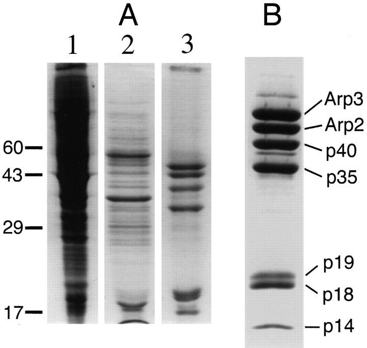
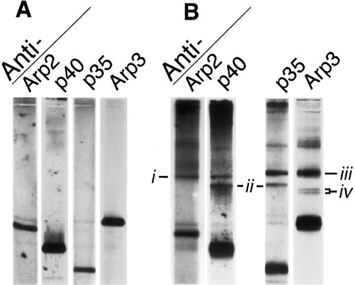
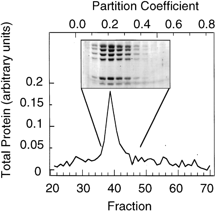
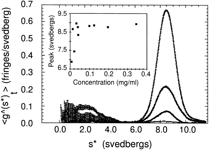
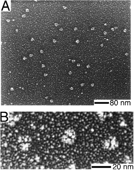
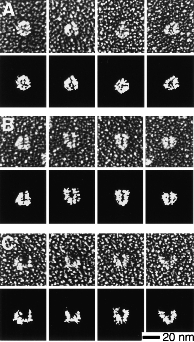
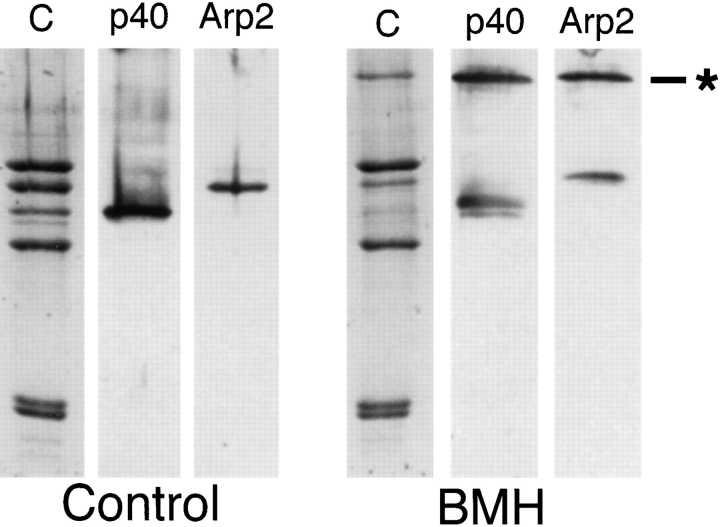
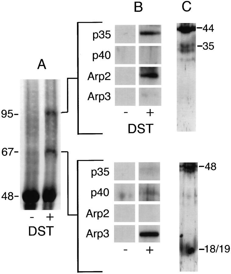
The Arp2/3 complex, first isolated from Acanthamoeba castellani by affinity chromatography on profilin, consists of seven polypeptides; two actin-related proteins, Arp2 and Arp3; and five apparently novel proteins, p40, p35, p19, p18, and p14 (Machesky et al., 1994). The complex is homogeneous by hydrodynamic criteria with a Stokes' radius of 5.3 nm by gel filtration, sedimentation coefficient of 8.7 S, and molecular mass of 197 kD by analytical ultracentrifugation. The stoichiometry of the subunits is 1:1:1:1:1:1:1, indicating the purified complex contains one copy each of seven polypeptides. In electron micrographs, the complex has a bilobed or horseshoe shape with outer dimensions of approximately 13 x 10 nm, and mathematical models of such a shape and size are consistent with the measured hydrodynamic properties. Chemical cross-linking with a battery of cross-linkers of different spacer arm lengths and chemical reactivities identify the following nearest neighbors within the complex: Arp2 and p40; Arp2 and p35; Arp3 and p35; Arp3 and either p18 or p19; and p19 and p14. By fluorescent antibody staining with anti-p40 and -p35, the complex is concentrated in the cortex of the ameba, especially in linear structures, possibly actin filament bundles, that lie perpendicular to the leading edge. Purified Arp2/3 complex binds actin filaments with a Kd of 2.3 microM and a stoichiometry of approximately one complex molecule per actin monomer. In electron micrographs of negatively stained samples, Arp2/3 complex decorates the sides of actin filaments. EDC/NHS cross-links actin to Arp3, p35, and a low molecular weight subunit, p19, p18, or p14. We propose structural and topological models for the Arp2/3 complex and suggest that affinity for actin filaments accounts for the localization of complex subunits to actin-rich regions of Acanthamoeba.
Arp2/3复合物最初是通过在肌动蛋白结合蛋白上进行亲和层析从卡氏棘阿米巴中分离出来的,它由七种多肽组成;两种肌动蛋白相关蛋白,Arp2和Arp3;以及五种明显的新蛋白,p40、p35、p19、p18和p14(马切斯基等人,1994年)。根据流体动力学标准,该复合物是均匀的,通过凝胶过滤法测得其斯托克斯半径为5.3纳米,沉降系数为8.7 S,通过分析超速离心法测得分子量为197 kD。亚基的化学计量比为1:1:1:1:1:1:1,表明纯化后的复合物包含七种多肽各一个拷贝。在电子显微镜照片中,该复合物呈双叶形或马蹄形,外部尺寸约为13×10纳米,这种形状和大小的数学模型与测得的流体动力学性质一致。用一系列具有不同间隔臂长度和化学反应性的交联剂进行化学交联,确定了复合物内以下最近邻关系:Arp2和p40;Arp2和p35;Arp3和p35;Arp3和p18或p19;以及p19和p14。通过用抗p40和-p35的荧光抗体染色,该复合物集中在阿米巴的皮质中,特别是在线性结构中,可能是与前缘垂直的肌动蛋白丝束中。纯化的Arp2/3复合物以2.3 microM的解离常数和每肌动蛋白单体约一个复合物分子的化学计量比结合肌动蛋白丝。在负染样品的电子显微镜照片中,Arp2/3复合物装饰在肌动蛋白丝的侧面。EDC/NHS将肌动蛋白与Arp3、p35以及一个低分子量亚基p19、p18或p14交联。我们提出了Arp2/3复合物的结构和拓扑模型,并表明对肌动蛋白丝的亲和力解释了复合物亚基在卡氏棘阿米巴富含肌动蛋白区域的定位。