Hoock T C, Peters L L, Lux S E
Division of Hematology/Oncology, Children's Hospital and the Dana Farber Cancer Institute, Boston, Massachusetts 02115, USA.
J Cell Biol. 1997 Mar 10;136(5):1059-70. doi: 10.1083/jcb.136.5.1059.
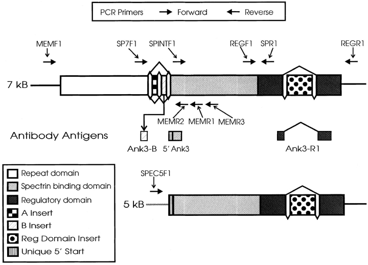
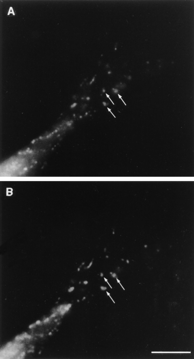
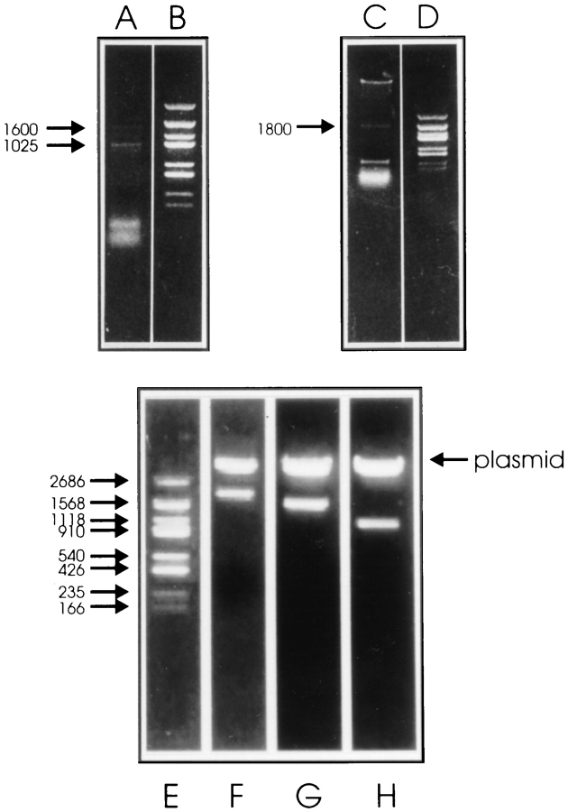
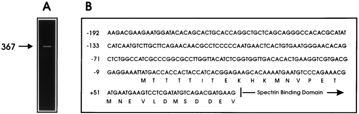
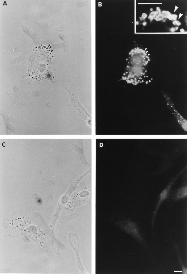
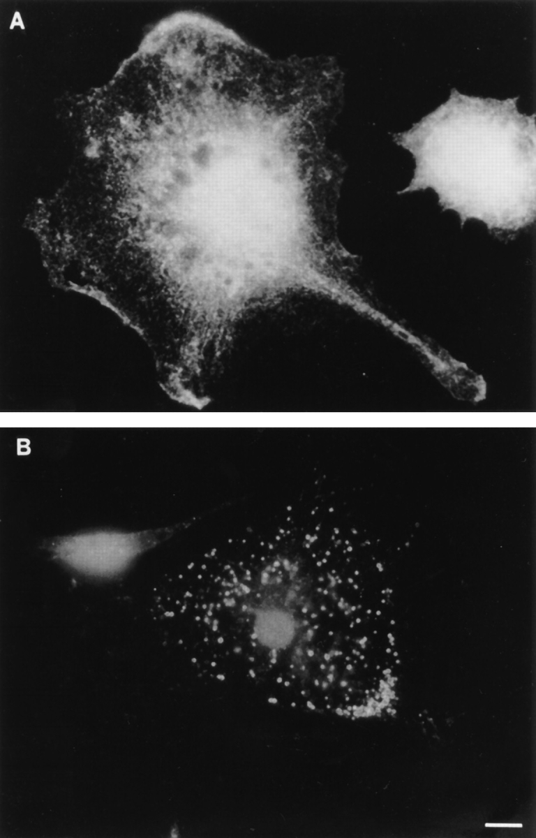
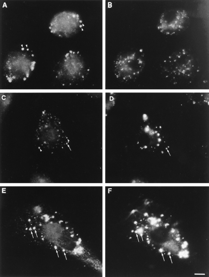
We have recently cloned and characterized ankyrin-3 (also called ankyrin(G)), a new ankyrin that is widely distributed, especially in epithelial tissues, muscle, and neuronal axons (Peters, L.L., K.M. John, F.M. Lu, E.M. Eicher, A. Higgins, M. Yialamas, L.C. Turtzo, A.J. Otsuka, and S.E. Lux. 1995. J. Cell Biol. 130: 313-330). Here we show that in mouse macrophages, ankyrin-3 is expressed exclusively as two small isoforms (120 and 100 kD) that lack the NH2-terminal repeats. Sequence analysis of isolated Ank3 cDNA clones, obtained by reverse transcription and amplification of mouse macrophage RNA (GenBank Nos. U89274 and U89275), reveals spectrin-binding and regulatory domains identical to those in kidney ankyrin-3 (GenBank No. L40631) preceded by a 29-amino acid segment of the membrane ("repeat") domain, beginning near the end of the last repeat. Antibodies specific for the regulatory and spectrin-binding domains of ankyrin-3 localize the protein to the surface of intracellular vesicles throughout the macrophage cytoplasm. It is not found on the plasma membrane. Also, epitope-tagged mouse macrophage ankyrin-3, transiently expressed in COS cells, associates with intracellular, not plasma, membranes. In contrast, ankyrin-1 (erythrocyte ankyrin, ankyrin(R)), which is also expressed in mouse macrophages, is located exclusively on the plasma membrane. The ankyrin-3-positive vesicles appear dark on phase-contrast microscopy. Two observations suggest that they are lysosomes. First, they are a late compartment in the endocytic pathway. They are only accessible to a fluorescent endocytic tracer (FITC-dextran) after a 24-h incubation, at which time all of the FITC-dextran-containing vesicles contain ankyrin-3 and vice versa. Second, the ankyrin-3-positive vesicles contain lysosomal-associated membrane glycoprotein (LAMP-1), a recognized lysosomal marker. This is the first evidence for the association of an ankyrin with lysosomes and is an example of two ankyrins present in the same cell that segregate to different locations.
我们最近克隆并鉴定了锚蛋白3(也称为锚蛋白(G)),这是一种新的锚蛋白,广泛分布,尤其在上皮组织、肌肉和神经元轴突中(彼得斯,L.L.,K.M.约翰,F.M.卢,E.M.艾歇尔,A.希金斯,M.亚拉马斯,L.C.图尔佐,A.J.大冢,和S.E.勒克斯。1995年。《细胞生物学杂志》130:313 - 330)。在此我们表明,在小鼠巨噬细胞中,锚蛋白3仅以两种缺少NH2末端重复序列的小异构体(120和100kD)形式表达。通过逆转录和扩增小鼠巨噬细胞RNA获得的分离的Ank3 cDNA克隆(基因库编号U89274和U89275)的序列分析显示,血影蛋白结合和调节结构域与肾锚蛋白3(基因库编号L40631)中的相同,其前面是膜(“重复”)结构域的一个29个氨基酸的片段,起始于最后一个重复序列末端附近。针对锚蛋白3的调节和血影蛋白结合结构域的特异性抗体将该蛋白定位到整个巨噬细胞胞质中的细胞内小泡表面。在质膜上未发现它。此外,在COS细胞中瞬时表达的表位标记的小鼠巨噬细胞锚蛋白3与细胞内而非质膜相关。相比之下,同样在小鼠巨噬细胞中表达的锚蛋白1(红细胞锚蛋白,锚蛋白(R))仅位于质膜上。在相差显微镜下,锚蛋白3阳性的小泡显得较暗。两项观察结果表明它们是溶酶体。第一,它们是内吞途径中的晚期区室。在24小时孵育后,荧光内吞示踪剂(FITC - 葡聚糖)才能进入它们,此时所有含FITC - 葡聚糖的小泡都含有锚蛋白3,反之亦然。第二,锚蛋白3阳性的小泡含有溶酶体相关膜糖蛋白(LAMP - 1),一种公认的溶酶体标记物。这是锚蛋白与溶酶体相关的首个证据,也是同一细胞中存在的两种锚蛋白分隔到不同位置的一个例子。