• 文献检索
  • 文档翻译
  • 深度研究
  • 学术资讯
  • Suppr Zotero 插件Zotero 插件
  • 邀请有礼
  • 套餐&价格
  • 历史记录
应用&插件
Suppr Zotero 插件Zotero 插件浏览器插件Mac 客户端Windows 客户端微信小程序
定价
高级版会员购买积分包购买API积分包
服务
文献检索文档翻译深度研究API 文档MCP 服务
关于我们
关于 Suppr公司介绍联系我们用户协议隐私条款
关注我们

Suppr 超能文献

核心技术专利:CN118964589B侵权必究
粤ICP备2023148730 号-1Suppr @ 2026

文献检索

告别复杂PubMed语法,用中文像聊天一样搜索,搜遍4000万医学文献。AI智能推荐,让科研检索更轻松。

立即免费搜索

文件翻译

保留排版,准确专业,支持PDF/Word/PPT等文件格式,支持 12+语言互译。

免费翻译文档

深度研究

AI帮你快速写综述,25分钟生成高质量综述,智能提取关键信息,辅助科研写作。

立即免费体验

人类腹主动脉瘤中膜退变时血管平滑肌细胞密度降低。

Decreased vascular smooth muscle cell density in medial degeneration of human abdominal aortic aneurysms.

作者信息

López-Candales A, Holmes D R, Liao S, Scott M J, Wickline S A, Thompson R W

机构信息

Division of Cardiology, Washington University School of Medicine, St. Louis, Missouri, USA.

出版信息

Am J Pathol. 1997 Mar;150(3):993-1007.

PMID:9060837
原文链接:https://pmc.ncbi.nlm.nih.gov/articles/PMC1857880/
Abstract

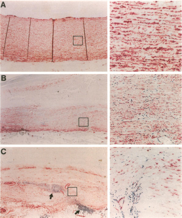
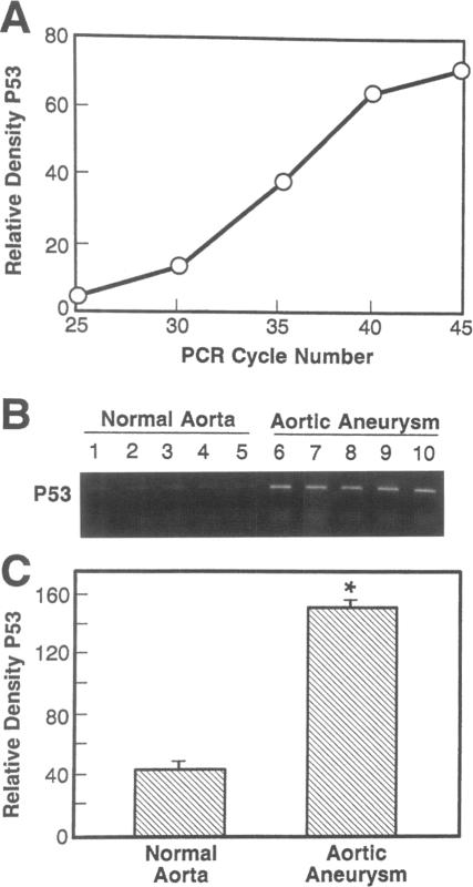
Abdominal aortic aneurysms (AAAs) are characterized by structural deterioration of the aortic wall leading to progressive aortic dilatation and eventual rupture. The histopathological changes in AAAs are particularly evident within the elastic media, which is normally dominated by vascular smooth muscle cells (SMCs). To determine whether a decrease in vascular SMCs contributes to medial degeneration, we measured SMC density in 21 normal and pathological human abdominal aortic tissue specimens using immunohistochemistry for alpha-SMC actin and direct cell counts (medial SMCs per high-power field (HPF)). Medial SMC density was not significantly different between normal aorta (n = 5; 199.5 +/- 14.9 SMCs/HPF) and atherosclerotic occlusive disease (n = 6; 176.4 +/- 13.9 SMCs/HPF), but it was reduced by 74% in AAA (n = 10; 50.9 +/- 6.1 SMCs/HPF; P < 0.01 versus normal aorta). Light and electron microscopy revealed no evidence of overt cellular necrosis, but SMCs in AAAs exhibited ultrastructural changes consistent with apoptosis. Using in situ end-labeling (ISEL) of fragmented DNA to detect apoptotic cells, up to 30% of aortic wall cells were ISEL positive in AAAs. By double-labeling techniques, many of these cells were alpha-actin-positive SMCs distributed throughout the degenerative media. In contrast, ISEL-positive cells were observed only within the intimal plaque in atherosclerotic occlusive disease. The amount of p53 protein detected by immunoblotting was increased nearly fourfold in AAA compared with normal aorta and atherosclerotic occlusive disease (P < 0.01), and immunoreactive p53 was localized to lymphocytes and residual SMCs in the aneurysm wall. Using reverse transcription polymerase chain reaction assays a substantial amount of p53 mRNA expression was observed in AAAs. These results demonstrate that medial SMC density is significantly decreased in human AAA tissues associated with evidence of SMC apoptosis and increased production of p53, a potential mediator of cell cycle arrest and programmed cell death. Given the role that SMCs normally play in maintaining medial architecture and in arterial wall matrix remodeling, the induction of SMC apoptosis likely makes an important contribution to the evolution of aneurysm degeneration.

摘要

腹主动脉瘤(AAA)的特征是主动脉壁结构退化,导致主动脉逐渐扩张并最终破裂。AAA的组织病理学变化在弹性中膜尤为明显,弹性中膜通常由血管平滑肌细胞(SMC)主导。为了确定血管SMC数量的减少是否导致中膜退变,我们使用α-SMC肌动蛋白免疫组织化学和直接细胞计数(每高倍视野(HPF)的中膜SMC数量)测量了21例正常和病理状态下的人类腹主动脉组织标本中的SMC密度。正常主动脉(n = 5;199.5±14.9个SMC/HPF)和动脉粥样硬化闭塞性疾病(n = 6;176.4±13.9个SMC/HPF)之间的中膜SMC密度无显著差异,但AAA组(n = 10;50.9±6.1个SMC/HPF;与正常主动脉相比,P < 0.01)降低了74%。光镜和电镜检查未发现明显的细胞坏死证据,但AAA中的SMC表现出与凋亡一致的超微结构变化。使用原位末端标记(ISEL)法检测断裂DNA以检测凋亡细胞,AAA中高达30%的主动脉壁细胞ISEL呈阳性。通过双重标记技术,这些细胞中有许多是分布在整个退变中膜的α-肌动蛋白阳性SMC。相比之下,在动脉粥样硬化闭塞性疾病中,仅在内膜斑块内观察到ISEL阳性细胞。免疫印迹法检测到的p53蛋白量在AAA中比正常主动脉和动脉粥样硬化闭塞性疾病增加了近四倍(P < 0.01),免疫反应性p53定位于动脉瘤壁中的淋巴细胞和残留的SMC。使用逆转录聚合酶链反应分析在AAA中观察到大量p53 mRNA表达。这些结果表明,在与SMC凋亡证据和p53产生增加相关的人类AAA组织中,中膜SMC密度显著降低,p53是细胞周期停滞和程序性细胞死亡的潜在介质。鉴于SMC通常在维持中膜结构和动脉壁基质重塑中所起的作用,SMC凋亡的诱导可能对动脉瘤退变的进展起重要作用。

https://cdn.ncbi.nlm.nih.gov/pmc/blobs/e3b2/1857880/6f54b858b236/amjpathol00027-0219-a.jpg
https://cdn.ncbi.nlm.nih.gov/pmc/blobs/e3b2/1857880/d2f6fea212ab/amjpathol00027-0214-a.jpg
https://cdn.ncbi.nlm.nih.gov/pmc/blobs/e3b2/1857880/79e9c3d24bd7/amjpathol00027-0216-a.jpg
https://cdn.ncbi.nlm.nih.gov/pmc/blobs/e3b2/1857880/8d2db4ee08f8/amjpathol00027-0217-a.jpg
https://cdn.ncbi.nlm.nih.gov/pmc/blobs/e3b2/1857880/ee78302f0127/amjpathol00027-0218-a.jpg
https://cdn.ncbi.nlm.nih.gov/pmc/blobs/e3b2/1857880/dbea692c46e7/amjpathol00027-0218-b.jpg
https://cdn.ncbi.nlm.nih.gov/pmc/blobs/e3b2/1857880/6f54b858b236/amjpathol00027-0219-a.jpg

相似文献

1
Decreased vascular smooth muscle cell density in medial degeneration of human abdominal aortic aneurysms.人类腹主动脉瘤中膜退变时血管平滑肌细胞密度降低。
Am J Pathol. 1997 Mar;150(3):993-1007.
2
Increased apoptosis and decreased density of medial smooth muscle cells in human abdominal aortic aneurysms.人腹主动脉瘤中内侧平滑肌细胞凋亡增加及密度降低。
Chin Med J (Engl). 2003 Oct;116(10):1549-52.
3
Death of smooth muscle cells and expression of mediators of apoptosis by T lymphocytes in human abdominal aortic aneurysms.人腹主动脉瘤中平滑肌细胞死亡及T淋巴细胞凋亡介质的表达
Circulation. 1999;99(1):96-104. doi: 10.1161/01.cir.99.1.96.
4
Comparison of cell-type-specific vs transmural aortic gene expression in experimental aneurysms.实验性动脉瘤中细胞类型特异性与跨壁主动脉基因表达的比较。
J Vasc Surg. 2005 May;41(5):844-52. doi: 10.1016/j.jvs.2005.02.027.
5
Enhanced embryonic nonmuscle myosin heavy chain isoform and matrix metalloproteinase expression in aortic abdominal aneurysm with rapid progression.进展迅速的腹主动脉瘤中胚胎非肌肉肌球蛋白重链亚型和基质金属蛋白酶表达增强。
Cardiovasc Pathol. 1999 Sep-Oct;8(5):291-5. doi: 10.1016/s1054-8807(99)00014-9.
6
Vascular smooth muscle cell apoptosis in aneurysmal, occlusive, and normal human aortas.人类动脉瘤性、闭塞性和正常主动脉中的血管平滑肌细胞凋亡
J Vasc Surg. 2000 Mar;31(3):567-76.
7
Receptor-interacting protein kinase 3 contributes to abdominal aortic aneurysms via smooth muscle cell necrosis and inflammation.受体相互作用蛋白激酶3通过平滑肌细胞坏死和炎症促进腹主动脉瘤的发生。
Circ Res. 2015 Feb 13;116(4):600-11. doi: 10.1161/CIRCRESAHA.116.304899. Epub 2015 Jan 6.
8
Accelerated replicative senescence of medial smooth muscle cells derived from abdominal aortic aneurysms compared to the adjacent inferior mesenteric artery.与相邻的肠系膜下动脉相比,腹主动脉瘤来源的中膜平滑肌细胞加速复制性衰老。
J Surg Res. 2000 Jul;92(1):85-95. doi: 10.1006/jsre.2000.5878.
9
Increased synthesis of matrix metalloproteinases by aortic smooth muscle cells is implicated in the etiopathogenesis of abdominal aortic aneurysms.主动脉平滑肌细胞中基质金属蛋白酶合成增加与腹主动脉瘤的发病机制有关。
J Vasc Surg. 1996 Jul;24(1):82-92. doi: 10.1016/s0741-5214(96)70148-9.
10
Medial neovascularization in abdominal aortic aneurysms: a histopathologic marker of aneurysmal degeneration with pathophysiologic implications.腹主动脉瘤的内侧新生血管形成:具有病理生理意义的动脉瘤退变的组织病理学标志物。
J Vasc Surg. 1995 May;21(5):761-71; discussion 771-2. doi: 10.1016/s0741-5214(05)80007-2.

引用本文的文献

1
A continuum model for tissues with moderate cell density.一种适用于中等细胞密度组织的连续体模型。
Comput Biol Med. 2025 Aug 30;197(Pt A):111025. doi: 10.1016/j.compbiomed.2025.111025.
2
The Pathophysiological Role of Vascular Smooth Muscle Cells in Abdominal Aortic Aneurysm.血管平滑肌细胞在腹主动脉瘤中的病理生理作用
Cells. 2025 Jul 2;14(13):1009. doi: 10.3390/cells14131009.
3
Single-cell RNA sequencing identifies two fibroblast subtypes and a Trem2 macrophage subtype as the possible specific cellular targets in abdominal aortic aneurysms.

本文引用的文献

1
Basic science of abdominal aortic aneurysms: emerging therapeutic strategies for an unresolved clinical problem.腹主动脉瘤的基础科学:针对一个尚未解决的临床问题的新兴治疗策略
Curr Opin Cardiol. 1996 Sep;11(5):504-18. doi: 10.1097/00001573-199609000-00010.
2
Vasoactive substances regulate vascular smooth muscle cell apoptosis. Countervailing influences of nitric oxide and angiotensin II.血管活性物质调节血管平滑肌细胞凋亡。一氧化氮和血管紧张素II的相反作用。
Circ Res. 1996 Oct;79(4):748-56. doi: 10.1161/01.res.79.4.748.
3
Contrary effects of lightly and strongly oxidized LDL with potent promotion of growth versus apoptosis on arterial smooth muscle cells, macrophages, and fibroblasts.
单细胞RNA测序确定了两种成纤维细胞亚型和一种Trem2巨噬细胞亚型,它们可能是腹主动脉瘤中特定的细胞靶点。
Front Immunol. 2025 May 20;16:1551308. doi: 10.3389/fimmu.2025.1551308. eCollection 2025.
4
Deficiency of IL-7R attenuates abdominal aortic aneurysms in mice by inhibiting macrophage polarization towards M1 phenotype through the NF-κB pathway.白细胞介素-7受体缺陷通过NF-κB途径抑制巨噬细胞向M1表型极化,从而减轻小鼠腹主动脉瘤。
Mol Med. 2025 Apr 16;31(1):138. doi: 10.1186/s10020-025-01209-2.
5
Myofibrillogenesis Regulator-1 in Smooth Muscle Cells Modulates Inflammation Signaling Pathways via Regulating ROCK1 Ubiquitination and Degradation to Impact Aortic Dissection.平滑肌细胞中的肌原纤维生成调节因子-1通过调节ROCK1泛素化和降解来调节炎症信号通路,从而影响主动脉夹层。
J Inflamm Res. 2025 Feb 5;18:1719-1738. doi: 10.2147/JIR.S485163. eCollection 2025.
6
Deficiency of ATF3 facilitates both angiotensin II-induced and spontaneously formed aortic aneurysm and dissection development by activating cGAS-STING pathway.ATF3的缺乏通过激活cGAS-STING通路促进血管紧张素II诱导的和自发形成的主动脉瘤及夹层的发展。
Clin Transl Med. 2025 Jan;15(1):e70147. doi: 10.1002/ctm2.70147.
7
Angiopoietin-2 reverses endothelial cell dysfunction in progeria vasculature.血管生成素-2可逆转早衰症血管中的内皮细胞功能障碍。
Aging Cell. 2025 Feb;24(2):e14375. doi: 10.1111/acel.14375. Epub 2024 Oct 18.
8
Endoplasmic reticulum stress in abdominal aortic aneurysm.腹主动脉瘤中的内质网应激
Int J Cardiol Heart Vasc. 2024 Aug 29;54:101500. doi: 10.1016/j.ijcha.2024.101500. eCollection 2024 Oct.
9
Coding and Non-Coding Transcriptomic Landscape of Aortic Complications in Marfan Syndrome.马凡综合征主动脉并发症的编码和非编码转录组景观。
Int J Mol Sci. 2024 Jul 5;25(13):7367. doi: 10.3390/ijms25137367.
10
The role of transcription factors in the pathogenesis and therapeutic targeting of vascular diseases.转录因子在血管疾病发病机制及治疗靶点中的作用。
Front Cardiovasc Med. 2024 Apr 30;11:1384294. doi: 10.3389/fcvm.2024.1384294. eCollection 2024.
轻度和重度氧化低密度脂蛋白对动脉平滑肌细胞、巨噬细胞和成纤维细胞具有相反的作用,前者有力地促进生长,而后者则促进凋亡。
Arterioscler Thromb Vasc Biol. 1996 Mar;16(3):416-24. doi: 10.1161/01.atv.16.3.416.
4
Differentiated vascular myocytes: are they involved in neointimal formation?分化的血管平滑肌细胞:它们参与新生内膜形成吗?
J Clin Invest. 1996 Feb 1;97(3):814-25. doi: 10.1172/JCI118481.
5
Apoptosis of vascular smooth muscle cells induced by in vitro stimulation with interferon-gamma, tumor necrosis factor-alpha, and interleukin-1 beta.γ干扰素、肿瘤坏死因子-α和白细胞介素-1β体外刺激诱导血管平滑肌细胞凋亡
Arterioscler Thromb Vasc Biol. 1996 Jan;16(1):19-27. doi: 10.1161/01.atv.16.1.19.
6
Sequencing of cDNA from 50 unrelated patients reveals that mutations in the triple-helical domain of type III procollagen are an infrequent cause of aortic aneurysms.对50名无亲缘关系患者的cDNA进行测序后发现,III型前胶原三螺旋结构域的突变是主动脉瘤的罕见病因。
J Clin Invest. 1993 Jun;91(6):2539-45. doi: 10.1172/JCI116490.
7
Enhanced production of the chemotactic cytokines interleukin-8 and monocyte chemoattractant protein-1 in human abdominal aortic aneurysms.人腹主动脉瘤中趋化细胞因子白细胞介素-8和单核细胞趋化蛋白-1的产生增加。
Am J Pathol. 1993 May;142(5):1423-31.
8
Thymocyte apoptosis induced by p53-dependent and independent pathways.由p53依赖和非依赖途径诱导的胸腺细胞凋亡。
Nature. 1993 Apr 29;362(6423):849-52. doi: 10.1038/362849a0.
9
Abdominal aortic aneurysm.腹主动脉瘤
N Engl J Med. 1993 Apr 22;328(16):1167-72. doi: 10.1056/NEJM199304223281607.
10
Adventitial elastolysis is a primary event in aneurysm formation.外膜弹性纤维溶解是动脉瘤形成的主要事件。
J Vasc Surg. 1993 Feb;17(2):371-80; discussion 380-1. doi: 10.1067/mva.1993.43023.