Carlevaro M F, Albini A, Ribatti D, Gentili C, Benelli R, Cermelli S, Cancedda R, Cancedda F D
Istituto Nazionale per la Ricerca sul Cancro, Università di Genova, Italy.
J Cell Biol. 1997 Mar 24;136(6):1375-84. doi: 10.1083/jcb.136.6.1375.
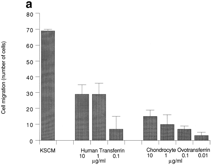
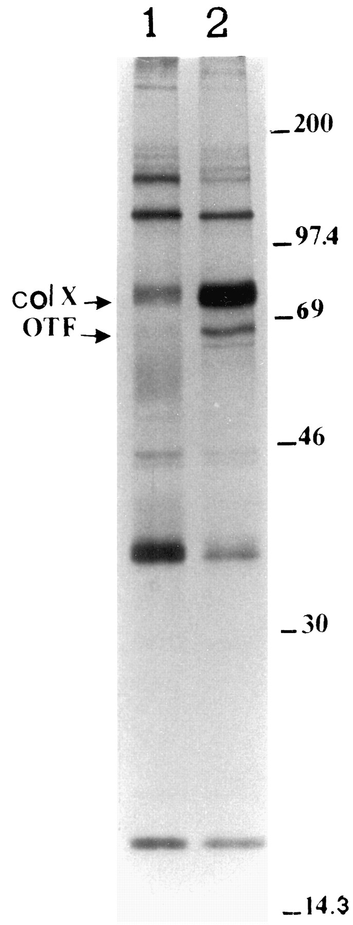
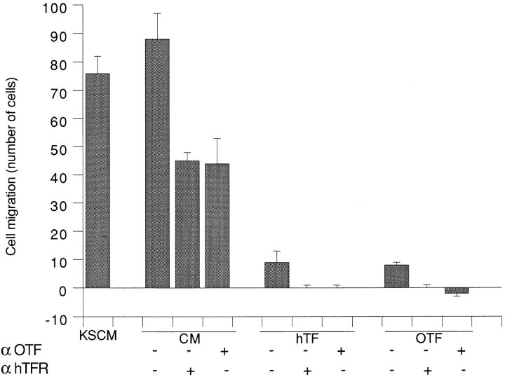
During endochondral bone formation, avascular cartilage differentiates to hypertrophic cartilage that then undergoes erosion and vascularization leading to bone deposition. Resting cartilage produces inhibitors of angiogenesis, shifting to production of angiogenic stimulators in hypertrophic cartilage. A major protein synthesized by hypertrophic cartilage both in vivo and in vitro is transferrin. Here we show that transferrin is a major angiogenic molecule released by hypertrophic cartilage. Endothelial cell migration and invasion is stimulated by transferrins from a number of different sources, including hypertrophic cartilage. Checkerboard analysis demonstrates that transferrin is a chemotactic and chemokinetic molecule. Chondrocyte-conditioned media show similar properties. Polyclonal anti-transferrin antibodies completely block endothelial cell migration and invasion induced by purified transferrin and inhibit the activity produced by hypertrophic chondrocytes by 50-70% as compared with controls. Function-blocking mAbs directed against the transferrin receptor similarly reduce the endothelial migratory response. Chondrocytes differentiating in the presence of serum produce transferrin, whereas those that differentiate in the absence of serum do not. Conditioned media from differentiated chondrocytes not producing transferrin have only 30% of the endothelial cell migratory activity of parallel cultures that synthesize transferrin. The angiogenic activity of transferrins was confirmed by in vivo assays on chicken egg chorioallantoic membrane, showing promotion of neovascularization by transferrins purified from different sources including conditioned culture medium. Based on the above results, we suggest that transferrin is a major angiogenic molecule produced by hypertrophic chondrocytes during endochondral bone formation.
在软骨内成骨过程中,无血管的软骨分化为肥大软骨,随后肥大软骨发生侵蚀和血管化,进而导致骨沉积。静止软骨产生血管生成抑制剂,而肥大软骨则转而产生血管生成刺激因子。肥大软骨在体内和体外合成的一种主要蛋白质是转铁蛋白。在此我们表明,转铁蛋白是肥大软骨释放的一种主要血管生成分子。来自多种不同来源(包括肥大软骨)的转铁蛋白可刺激内皮细胞迁移和侵袭。棋盘分析表明,转铁蛋白是一种趋化性和化学动力学分子。软骨细胞条件培养基具有类似特性。多克隆抗转铁蛋白抗体可完全阻断纯化转铁蛋白诱导的内皮细胞迁移和侵袭,并与对照组相比,将肥大软骨细胞产生的活性抑制50% - 70%。针对转铁蛋白受体的功能阻断单克隆抗体同样可降低内皮细胞迁移反应。在有血清存在的情况下分化的软骨细胞产生转铁蛋白,而在无血清情况下分化的软骨细胞则不产生。未产生转铁蛋白的分化软骨细胞的条件培养基,其内皮细胞迁移活性仅为合成转铁蛋白的平行培养物的30%。通过在鸡胚绒毛尿囊膜上进行的体内试验证实了转铁蛋白具有血管生成活性,结果显示从包括条件培养基在内的不同来源纯化的转铁蛋白可促进新血管形成。基于上述结果,我们认为转铁蛋白是肥大软骨细胞在软骨内成骨过程中产生的一种主要血管生成分子。