• 文献检索
  • 文档翻译
  • 深度研究
  • 学术资讯
  • Suppr Zotero 插件Zotero 插件
  • 邀请有礼
  • 套餐&价格
  • 历史记录
应用&插件
Suppr Zotero 插件Zotero 插件浏览器插件Mac 客户端Windows 客户端微信小程序
定价
高级版会员购买积分包购买API积分包
服务
文献检索文档翻译深度研究API 文档MCP 服务
关于我们
关于 Suppr公司介绍联系我们用户协议隐私条款
关注我们

Suppr 超能文献

核心技术专利:CN118964589B侵权必究
粤ICP备2023148730 号-1Suppr @ 2026

文献检索

告别复杂PubMed语法,用中文像聊天一样搜索,搜遍4000万医学文献。AI智能推荐,让科研检索更轻松。

立即免费搜索

文件翻译

保留排版,准确专业,支持PDF/Word/PPT等文件格式,支持 12+语言互译。

免费翻译文档

深度研究

AI帮你快速写综述,25分钟生成高质量综述,智能提取关键信息,辅助科研写作。

立即免费体验

鲍鱼壳蛋白对方解石晶体生长的修饰:原子力显微镜研究

Modification of calcite crystal growth by abalone shell proteins: an atomic force microscope study.

作者信息

Walters D A, Smith B L, Belcher A M, Paloczi G T, Stucky G D, Morse D E, Hansma P K

机构信息

Department of Physics, University of California, Santa Barbara 93106, USA.

出版信息

Biophys J. 1997 Mar;72(3):1425-33. doi: 10.1016/S0006-3495(97)78789-7.

DOI:10.1016/S0006-3495(97)78789-7
PMID:9138588
原文链接:https://pmc.ncbi.nlm.nih.gov/articles/PMC1184525/
Abstract

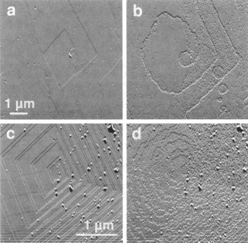
A family of soluble proteins from the shell of Haliotis rufescens was introduced over a growing calcite crystal being scanned in situ by an atomic force microscope (AFM). Atomic step edges on the crystal surface were altered in shape and speed of growth by the proteins. Proteins attached nonuniformly to the surface, indicating different interactions with crystallographically different step edges. The observed changes were consistent with the habit modification induced by this family of proteins, as previously observed by optical microscopy. To facilitate further studies in this area, AFM techniques and certain AFM imaging artifacts are discussed in detail.

摘要

将来自红鲍贝壳的一族可溶性蛋白质引入到一个正在通过原子力显微镜(AFM)进行原位扫描的方解石生长晶体上。晶体表面的原子台阶边缘在形状和生长速度上因这些蛋白质而发生改变。蛋白质在表面的附着不均匀,这表明与晶体学上不同的台阶边缘存在不同的相互作用。观察到的这些变化与此前通过光学显微镜观察到的由该族蛋白质诱导的习性改变是一致的。为便于在该领域开展进一步研究,详细讨论了AFM技术及某些AFM成像假象。

https://cdn.ncbi.nlm.nih.gov/pmc/blobs/fae1/1184525/2e2b8dbe2904/biophysj00036-0451-b.jpg
https://cdn.ncbi.nlm.nih.gov/pmc/blobs/fae1/1184525/05938f3477d3/biophysj00036-0446-a.jpg
https://cdn.ncbi.nlm.nih.gov/pmc/blobs/fae1/1184525/b5d20af0db0f/biophysj00036-0447-a.jpg
https://cdn.ncbi.nlm.nih.gov/pmc/blobs/fae1/1184525/454701fa9932/biophysj00036-0448-a.jpg
https://cdn.ncbi.nlm.nih.gov/pmc/blobs/fae1/1184525/cb82b5e95a58/biophysj00036-0449-a.jpg
https://cdn.ncbi.nlm.nih.gov/pmc/blobs/fae1/1184525/6ec2647d5c3b/biophysj00036-0449-b.jpg
https://cdn.ncbi.nlm.nih.gov/pmc/blobs/fae1/1184525/eb8b4d70e217/biophysj00036-0451-a.jpg
https://cdn.ncbi.nlm.nih.gov/pmc/blobs/fae1/1184525/2e2b8dbe2904/biophysj00036-0451-b.jpg

相似文献

1
Modification of calcite crystal growth by abalone shell proteins: an atomic force microscope study.鲍鱼壳蛋白对方解石晶体生长的修饰:原子力显微镜研究
Biophys J. 1997 Mar;72(3):1425-33. doi: 10.1016/S0006-3495(97)78789-7.
2
Direct observation of the transition from calcite to aragonite growth as induced by abalone shell proteins.直接观察鲍鱼壳蛋白诱导方解石向文石生长的转变过程。
Biophys J. 2000 Dec;79(6):3307-12. doi: 10.1016/S0006-3495(00)76562-3.
3
Scanning-induced growth on single crystal calcite with an atomic force microscope.利用原子力显微镜在方解石单晶上进行扫描诱导生长
Langmuir. 2006 Aug 1;22(16):6931-8. doi: 10.1021/la0608359.
4
Characterization of two molluscan crystal-modulating biomineralization proteins and identification of putative mineral binding domains.两种软体动物晶体调节生物矿化蛋白的表征及假定矿物结合结构域的鉴定
Biopolymers. 2003 Dec;70(4):522-33. doi: 10.1002/bip.10536.
5
Perlwapin, an abalone nacre protein with three four-disulfide core (whey acidic protein) domains, inhibits the growth of calcium carbonate crystals.珍珠层蛋白是一种具有三个四二硫键核心(乳清酸性蛋白)结构域的鲍鱼珍珠层蛋白,可抑制碳酸钙晶体的生长。
Biophys J. 2006 Oct 1;91(7):2601-8. doi: 10.1529/biophysj.106.086108. Epub 2006 Jul 21.
6
Perlinhibin, a cysteine-, histidine-, and arginine-rich miniprotein from abalone (Haliotis laevigata) nacre, inhibits in vitro calcium carbonate crystallization.珍珠抑制素是一种来自鲍鱼(皱纹盘鲍)珍珠层的富含半胱氨酸、组氨酸和精氨酸的小蛋白,它能在体外抑制碳酸钙结晶。
Biophys J. 2007 Aug 15;93(4):1246-54. doi: 10.1529/biophysj.106.100636. Epub 2007 May 11.
7
Self-Assembly of Protein Nanofibrils Orchestrates Calcite Step Movement through Selective Nonchiral Interactions.蛋白纳米原纤维的自组装通过选择性非手性相互作用来调控方解石阶跃运动。
ACS Nano. 2015 Jun 23;9(6):5782-91. doi: 10.1021/acsnano.5b01870. Epub 2015 May 20.
8
Formation of chiral morphologies through selective binding of amino acids to calcite surface steps.通过氨基酸与方解石表面台阶的选择性结合形成手性形态。
Nature. 2001 Jun 14;411(6839):775-9. doi: 10.1038/35081034.
9
Spatially resolved product formation in the reaction of formic acid with calcium carbonate (1014): the role of step density and adsorbed water-assisted ion mobility.
Langmuir. 2007 Jun 19;23(13):7039-45. doi: 10.1021/la062866+. Epub 2007 May 15.
10
The nacre protein perlucin nucleates growth of calcium carbonate crystals.珍珠层蛋白perlucin可促使碳酸钙晶体生长成核。
J Microsc. 2003 Dec;212(Pt 3):280-91. doi: 10.1111/j.1365-2818.2003.01263.x.

引用本文的文献

1
Exploring the Potential of Nonclassical Crystallization Pathways to Advance Cementitious Materials.探索非经典结晶途径在推进胶凝材料方面的潜力。
Chem Rev. 2024 Jun 26;124(12):7538-7618. doi: 10.1021/acs.chemrev.3c00259. Epub 2024 Jun 14.
2
In-depth proteomic analyses of (greenlip abalone) nacre and prismatic organic shell matrix.对(绿唇鲍)珍珠层和棱柱形有机贝壳基质进行深入的蛋白质组学分析。
Proteome Sci. 2018 Jun 15;16:11. doi: 10.1186/s12953-018-0139-3. eCollection 2018.
3
Effects of acidic peptide size and sequence on trivalent praseodymium adduction and electron transfer dissociation mass spectrometry.

本文引用的文献

1
Biological control of crystal texture: a widespread strategy for adapting crystal properties to function.生物控制晶体结构:一种广泛应用的策略,用于使晶体性质适应功能。
Science. 1993 Feb 5;259(5096):776-9. doi: 10.1126/science.259.5096.776.
2
Crystal tectonics: construction of reticulated calcium phosphate frameworks in bicontinuous reverse microemulsions.晶体构造:双连续反相胶束中网状磷酸钙骨架的构建。
Science. 1994 Jun 10;264(5165):1576-8. doi: 10.1126/science.264.5165.1576.
3
Mechanisms of protein crystal growth: An atomic force microscopy study of canavalin crystallization.
酸性肽的大小和序列对三价镨加合及电子转移解离质谱分析的影响
J Mass Spectrom. 2017 Apr;52(4):218-229. doi: 10.1002/jms.3919.
4
Exploiting fluorescence resonance energy transfer to probe structural changes in a macromolecule during adsorption and incorporation into a growing biomineral crystal.利用荧光共振能量转移技术探测在大分子吸附和整合到生长的生物矿化晶体过程中的结构变化。
Colloids Surf B Biointerfaces. 2009 Dec 1;74(2):401-9. doi: 10.1016/j.colsurfb.2009.07.004. Epub 2009 Jul 14.
5
Perlinhibin, a cysteine-, histidine-, and arginine-rich miniprotein from abalone (Haliotis laevigata) nacre, inhibits in vitro calcium carbonate crystallization.珍珠抑制素是一种来自鲍鱼(皱纹盘鲍)珍珠层的富含半胱氨酸、组氨酸和精氨酸的小蛋白,它能在体外抑制碳酸钙结晶。
Biophys J. 2007 Aug 15;93(4):1246-54. doi: 10.1529/biophysj.106.100636. Epub 2007 May 11.
6
Perlwapin, an abalone nacre protein with three four-disulfide core (whey acidic protein) domains, inhibits the growth of calcium carbonate crystals.珍珠层蛋白是一种具有三个四二硫键核心(乳清酸性蛋白)结构域的鲍鱼珍珠层蛋白,可抑制碳酸钙晶体的生长。
Biophys J. 2006 Oct 1;91(7):2601-8. doi: 10.1529/biophysj.106.086108. Epub 2006 Jul 21.
7
Mechanical properties of nacre and highly mineralized bone.珍珠母和高度矿化骨的力学性能。
Proc Biol Sci. 2001 Jan 7;268(1462):107-11. doi: 10.1098/rspb.2000.1337.
8
Direct observation of the transition from calcite to aragonite growth as induced by abalone shell proteins.直接观察鲍鱼壳蛋白诱导方解石向文石生长的转变过程。
Biophys J. 2000 Dec;79(6):3307-12. doi: 10.1016/S0006-3495(00)76562-3.
9
Cell-mediated crystallization of calcium oxalate in plants.植物中草酸钙的细胞介导结晶
Plant Cell. 1999 Apr;11(4):751-61. doi: 10.1105/tpc.11.4.751.
10
A peptide that inhibits hydroxyapatite growth is in an extended conformation on the crystal surface.一种抑制羟基磷灰石生长的肽在晶体表面呈伸展构象。
Proc Natl Acad Sci U S A. 1998 Oct 13;95(21):12083-7. doi: 10.1073/pnas.95.21.12083.
蛋白质晶体生长机制:刀豆球蛋白结晶的原子力显微镜研究
Phys Rev Lett. 1995 Oct 2;75(14):2774-2777. doi: 10.1103/PhysRevLett.75.2774.
4
Probing oscillatory hydration potentials using thermal-mechanical noise in an atomic-force microscope.
Phys Rev B Condens Matter. 1995 Sep 15;52(12):R8692-R8695. doi: 10.1103/physrevb.52.r8692.
5
Protein tracking and detection of protein motion using atomic force microscopy.使用原子力显微镜进行蛋白质追踪及蛋白质运动检测。
Biophys J. 1996 May;70(5):2421-31. doi: 10.1016/S0006-3495(96)79812-0.
6
Atomic force microscopy of insulin single crystals: direct visualization of molecules and crystal growth.胰岛素单晶的原子力显微镜:分子与晶体生长的直接可视化
Biophys J. 1996 Aug;71(2):1071-8. doi: 10.1016/S0006-3495(96)79307-4.
7
Atomic force microscopy and molecular modeling of protein and peptide binding to calcite.蛋白质和肽与方解石结合的原子力显微镜及分子模拟
Calcif Tissue Int. 1994 Feb;54(2):133-41. doi: 10.1007/BF00296064.
8
Urinary calcium oxalate crystal growth inhibitors.草酸钙尿结石晶体生长抑制剂。
J Am Soc Nephrol. 1994 Nov;5(5 Suppl 1):S46-53. doi: 10.1681/ASN.V55s46.
9
Analysis of the soluble organic matrix of five morphologically different kidney stones. Evidence for a specific role of albumin in the constitution of the stone protein matrix.五种形态各异肾结石的可溶性有机基质分析。白蛋白在结石蛋白基质构成中特定作用的证据。
Urol Res. 1995;23(1):45-51. doi: 10.1007/BF00298850.
10
Applications for atomic force microscopy of DNA.DNA的原子力显微镜应用。
Biophys J. 1995 May;68(5):1672-7. doi: 10.1016/S0006-3495(95)80343-7.