Zheng J, Rudra-Ganguly N, Miller G J, Moffatt K A, Cote R J, Roy-Burman P
Department of Pathology, University of Southern California School of Medicine, Los Angeles 90033, USA.
Am J Pathol. 1997 Jun;150(6):2009-18.
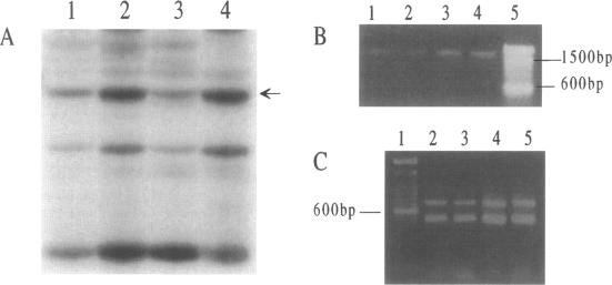
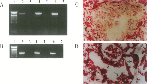
Application of differential display to the comparison of androgen-stimulated and unstimulated human prostate carcinoma cell line LNCaP identified androgen induction of the L-plastin gene, which encodes an actin-binding protein isoform. Further investigation demonstrated that L-plastin expression in LNCaP cells is up-regulated by both dihydrotestosterone and estradiol. This induction of expression is detected as early as 2 hours after addition of steroids to the cell culture. L-plastin expression is also detected in other prostate carcinoma cell lines by reverse transcriptase polymerase chain reaction and immunohistochemistry but not in the single normal adult prostate epithelial cell line that is available to us. Analysis of multiple primary prostatic tumor tissues as well as normal and tumor tissues of the same prostate gland showed that tumor tissues exhibit a higher level of expression as compared with the normal tissues. Immunohistochemical study using anti-L-plastin antiserum on normal and carcinomatous prostate tissues showed a very striking difference in the staining patterns. Positive staining was seen in the fibromuscular stroma in normal prostates but not in the glandular epithelial cells. In contrast, strong staining was seen predominantly within the carcinomatous glandular epithelial cells. Taken together, these results suggest that the expression of L-plastin in prostatic epithelial cells is linked to the malignant state and that, once expressed in carcinomas, its expression is regulated by steroid hormone receptors.
应用差异显示技术比较雄激素刺激和未刺激的人前列腺癌细胞系LNCaP,发现雄激素可诱导L-丝动蛋白基因表达,该基因编码一种肌动蛋白结合蛋白异构体。进一步研究表明,LNCaP细胞中L-丝动蛋白的表达受双氢睾酮和雌二醇上调。在细胞培养中加入类固醇后最早2小时就能检测到这种表达诱导。通过逆转录聚合酶链反应和免疫组织化学也在其他前列腺癌细胞系中检测到L-丝动蛋白表达,但在我们可获得的单一正常成人前列腺上皮细胞系中未检测到。对多个原发性前列腺肿瘤组织以及同一前列腺的正常和肿瘤组织分析表明,与正常组织相比,肿瘤组织呈现更高水平的表达。使用抗L-丝动蛋白抗血清对正常和癌性前列腺组织进行免疫组织化学研究,结果显示染色模式存在非常显著的差异。在正常前列腺的纤维肌基质中可见阳性染色,但在腺上皮细胞中未见。相反,在癌性腺上皮细胞中主要可见强染色。综上所述,这些结果表明L-丝动蛋白在前列腺上皮细胞中的表达与恶性状态相关,并且一旦在癌中表达,其表达受类固醇激素受体调节。